. 2021 Dec;125(12):1657-1665.
doi: 10.1038/s41416-021-01585-6. Epub 2021 Oct 20.

Preclinical study of 212Pb alpha-radioimmunotherapy targeting CD20 in non-Hodgkin lymphoma

Affiliations

Preclinical study of 212Pb alpha-radioimmunotherapy targeting CD20 in non-Hodgkin lymphoma

Stéphanie Durand-Panteix et al. Br J Cancer. 2021 Dec.

Abstract

Background: Despite therapeutic advances, Non-Hodgkin lymphoma (NHL) relapses can occur. The development of radioimmunotherapy (RIT) with α-emitters is an attractive alternative. In this study, we investigated the potential of α-RIT in conjunction with 212Pb-rituximab for the treatment of NHL.

Methods: EL4-hCD20-Luc cells (mouse lymphoma cell line) were used for in vitro and in vivo studies. Biodistribution and efficacy studies were performed on C57BL/6 mice injected intravenously with 25 × 103 cells.

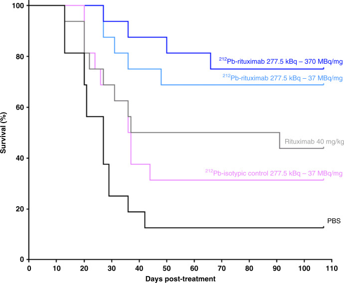
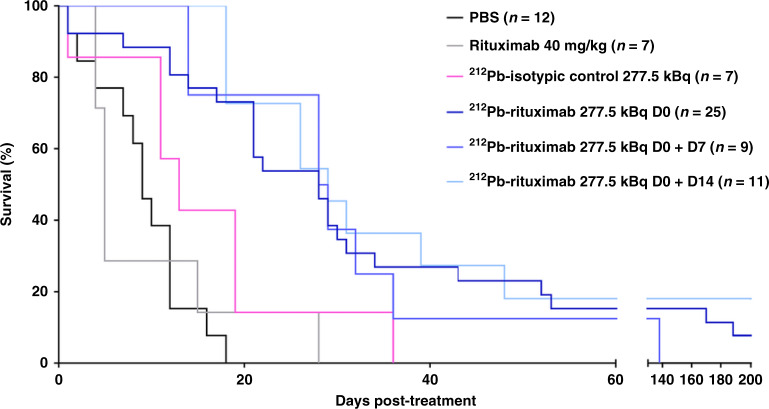
Results: 212Pb-rituximab (0.925-7.4 kBq/mL) inhibit proliferation of EL4-hCD20-Luc cells in vitro. Biodistribution of 203/212Pb-rituximab in mice showed a significant tumour uptake and suggested that the liver, spleen, and kidneys were the organs at risk. For efficacy studies, mice were treated at either 11 days (early stage) or 20-30 days after injection of tumour cells (late stage). Treatment with 277.5 kBq 212Pb-rituximab significantly prolonged survival. Even at an advanced tumour stage, significant tumour regression occurred, with an increase in the median survival time to 28 days, compared with 9 days in the controls.

Conclusions: These results show the efficacy of 212Pb-rituximab in a murine syngeneic lymphoma model, in terms of significant tumour regression and increased survival, thereby highlighting the potency of α-RIT for the treatment of NHL.


Conflict of interest statement

Amal Saidi and Julien Torgue are Orano Med employees. No other potential conflicts of interest relevant to this article exist.

Figures

Fig. 1
Fig. 1. In vitro studies.
Percentage of growth inhibition (a) and mortality (b) for increasing 212Pb-mAb activity (0.925–7.4 kBq/mL). a EL4-hCD20-luc cell growth inhibition percent was calculated, from days 1 to 3, using untreated cells as controls on the same day. Data are expressed as the mean ± SEM percentage of growth inhibition ((1 – ODtreatment /ODcontrol) * 100). b Mortality was evaluated on day 2 by flow cytometry using Annexin V/7AAD dual staining to evaluate early apoptotic, late apoptotic, and necrotic cells.
Fig. 2
Fig. 2. Biodistribution and SPECT-CT imaging.
Biodistribution of 212Pb-rituximab (a) and the 212Pb-isotypic control (b) in tumour-bearing mice. Mice were injected with 185 kBq 212Pb-mAbs and then sacrificed at 2, 6, 18, 24, or 48 h post-injection. Radioactivity in tumours, organs, and blood was expressed as the mean ± SD percentage injected dose per gram of tissue (%ID/g). In tumours, two-way analysis of variance showed a significant difference between the two mAbs (p < 0.006). In vivo imaging of 203Pb-rituximab in tumour-bearing mice (c) and healthy mice (d). 203Pb-rituximab at 18.5–25 MBq was injected. Mice were imaged by MicroSPECT/CT at 6, 24, 48, and 96 h after injection. For mice bearing tumours (25 days after engraftment), BLI was performed before injection of 203Pb-rituximab and after the last MicroSPECT/CT acquisition. Each line represents the same mouse imaged at different times. Tumours are indicated by different arrows, allowing monitoring of tumour uptake at the same site. Tumoral presence was confirmed post-mortem at the end of experiment.
Fig. 3
Fig. 3. Toxicity studies.
A Effect of 212Pb-rituximab on the survival of healthy mice. Three time points were evaluated: 7 days, 21 days, and 3 months post-injection. Values in bold indicate when lethality occurred. B 212Pb-rituximab toxicity studies based on haematological parameters. The white blood cell count (a), platelet count (b), and haemoglobin concentration (c) were measured at 7 days, 21 days, and 3 months post-injection of PBS, 212Pb-isotypic control (277.5 or 555 kBq), or 212Pb-rituximab (185–740 kBq). Data represent the mean ± SEM. *All mice died before the endpoint.
Fig. 4
Fig. 4. Efficacy of 212Pb-rituximab treatment in a murine syngeneic lymphoma model at an early stage.
At 11 days post-engraftment, mice received the following treatments (n = 16 per group): PBS, 40 mg/kg rituximab, 277.5 kBq 212Pb-isotypic control, and 277 kBq 212Pb-rituximab with a specific activity of 370 MBq/mg or 37 MBq/mg. Survival rates were determined by the Kaplan–Meier method and compared using the log-rank test. All treatments except the 212Pb-isotypic control induced a significant increase in MST compared with PBS.
Fig. 5
Fig. 5. Efficacy of 212Pb-rituximab treatment in a late-stage murine syngeneic lymphoma model.
Once the tumour was detected by BLI (20–30 days after cell injection), mice were treated with an injection of PBS, rituximab (40 mg/kg), 212Pb-isotypic control (277.5 kBq), 212Pb-rituximab (277.5 kBq), or two injections of 212Pb-rituximab (277.5 kBq) on days 0 and 7 or days 0 and 14. Survival rates were determined by the Kaplan–Meier method and compared using the log-rank test. All 212Pb-rituximab treatments induced a significant increase in the MST compared with PBS (p ≤ 0.001).
Fig. 6
Fig. 6. BLI monitoring in a late-stage murine syngeneic lymphoma model.
When the tumour was detected by BLI (20–30 days after cell injection), mice were injected with PBS, rituximab (40 mg/kg), 212Pb-isotypic control (277.5 kBq), 212Pb- rituximab (277.5 kBq) on day 0 (T0), or 212Pb-rituximab (277.5 kBq) on T0 and day 7 (T7) or T0 and day 14 (T14). Mice were monitored by BLI at the indicated time points to follow tumour evolution until sacrifice or the end of the experiment.

References

    1. Shankland KR, Armitage JO, Hancock BW. Non-Hodgkin lymphoma. Lancet. 2012;380:848–57. doi: 10.1016/S0140-6736(12)60605-9. - DOI - PubMed
    1. Beers SA, Chan CHT, French RR, Cragg MS, Glennie MJ. CD20 as a target for therapeutic type I and II monoclonal antibodies. Semin Hematol. 2010;47:107–14. doi: 10.1053/j.seminhematol.2010.01.001. - DOI - PubMed
    1. Witzig TE. Radioimmunotherapy for B-cell non-Hodgkin lymphoma. Best Pract Res Clin Haematol. 2006;19:655–68. doi: 10.1016/j.beha.2006.05.002. - DOI - PubMed
    1. Eskian M, Khorasanizadeh M, Zinzani PL, Illidge TM, Rezaei N. Novel methods to improve the efficiency of radioimmunotherapy for non-Hodgkin lymphoma. Int Rev Immunol. 2019;38:79–91. doi: 10.1080/08830185.2019.1588266. - DOI - PubMed
    1. Baidoo KE, Yong K, Brechbiel MW. Molecular pathways: targeted α-particle radiation therapy. Clin Cancer Res. 2013;19:530–7. doi: 10.1158/1078-0432.CCR-12-0298. - DOI - PMC - PubMed

Publication types