. 2021 Oct 4:12:750200.
doi: 10.3389/fmicb.2021.750200. eCollection 2021.

Monitoring Bacterial Conjugation by Optical Microscopy

Affiliations

Monitoring Bacterial Conjugation by Optical Microscopy

Gerardo Carranza et al. Front Microbiol. .

Abstract

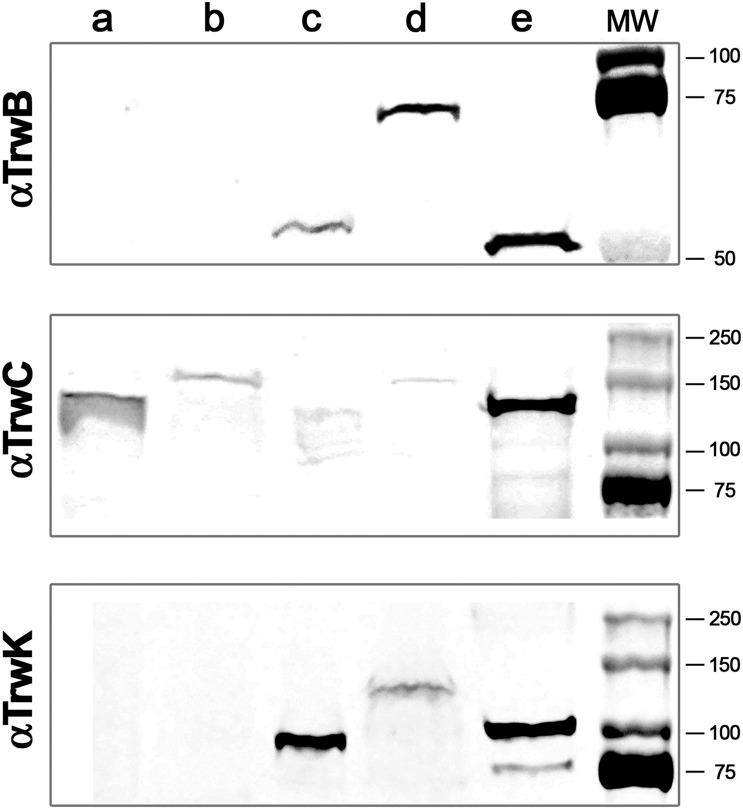
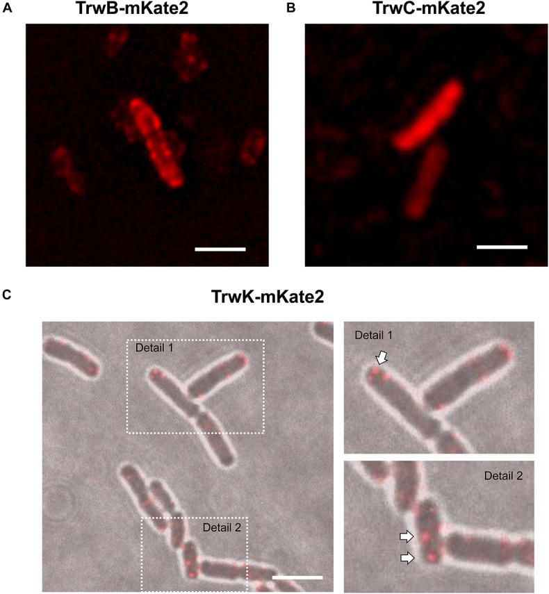
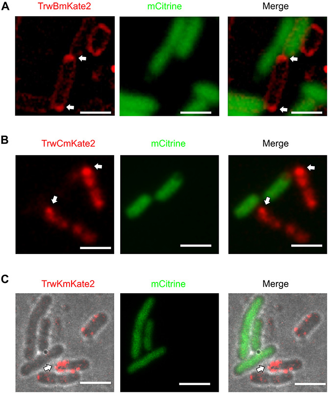
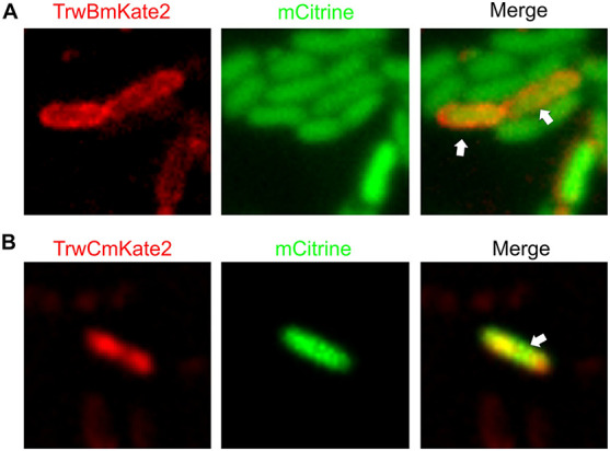
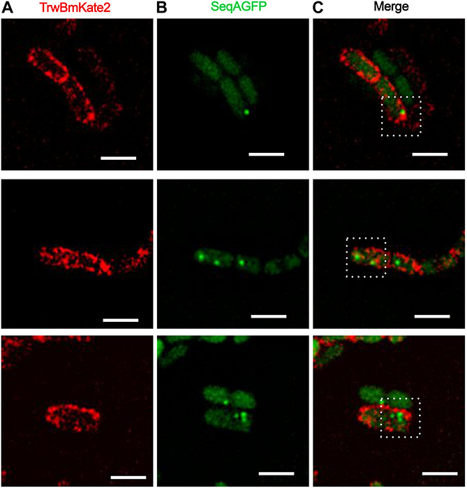
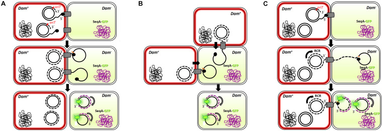
Bacterial conjugation is the main mechanism for horizontal gene transfer, conferring plasticity to the genome repertoire. This process is also the major instrument for the dissemination of antibiotic resistance genes. Hence, gathering primary information of the mechanism underlying this genetic transaction is of a capital interest. By using fluorescent protein fusions to the ATPases that power conjugation, we have been able to track the localization of these proteins in the presence and absence of recipient cells. Moreover, we have found that more than one copy of the conjugative plasmid is transferred during mating. Altogether, these findings provide new insights into the mechanism of such an important gene transfer device.

Keywords: T4SS; antibiotic resistance; bacterial conjugation; conjugative ATPases; fluorescence microscopy.


Conflict of interest statement

The authors declare that the research was conducted in the absence of any commercial or financial relationships that could be construed as a potential conflict of interest.

Figures

FIGURE 1
FIGURE 1
Immunodetection and subcellular localization of conjugative ATPases. MG1655 Escherichia coli cells were transformed with R388 wild type plasmid or with R388 encoding for TrwBmKate2, TrwCmKate2, or TrwKmKate2. Cells were grown to stationary phase and harvested by centrifugation. Cell pellets were re-suspended and lysed by mechanical methods. Soluble and membrane fractions were separated by ultra-centrifugation. Aliquots of each fraction were run in a SDS-PAGE. Gels were transferred to a nitrocellulose membrane and incubated with antibodies anti-TrwB (upper panel), anti-TrwC (middle panel), or anti-TrwK (bottom panel) and then revealed with IRDye. Upper panel: (a) wild type R388 soluble fraction, (b) R388_TrwBmKate2 soluble fraction, (c) wild type R388 membrane fraction, (d) R388_TrwBmKate2 membrane fraction, and (e) purified TrwBΔN70 protein. Middle panel: (a) wild type R388 soluble fraction, (b) R388_TrwCmKate2 soluble fraction, (c) wild type R388 membrane fraction, (d) R388_TrwCmKate2 membrane fraction, and (e) purified TrwC protein. Bottom panel: (a) wild type R388 soluble fraction, (b) R388_TrwKmKate2 soluble fraction, (c) wild type R388 membrane fraction, (d) R388_TrwKmKate2 membrane fraction, and (e) purified TrwK protein. Estimated MW values for TrwB, TrwBmKate2, and TrwBΔN70 proteins are 56.3, 82.4, and 48.6 kDa, respectively. Estimated MW values for TrwC and TrwCmKate2 proteins are 107.4 and 133.5 kDa, respectively. Estimated MW values for TrwK and TrwKmKate2 proteins are 93.8 and 119.9 kDa, respectively.
FIGURE 2
FIGURE 2
Localization of conjugative proteins in donor cells in the absence of recipients. MG1655 cells expressing TrwBmKate2 (A), TrwCmKate2 (B), and TrwKmKate2 (C) were grown in M9 minimal media and examined by fluorescent microscopy. In all cases, fusion proteins are expressed under its respective natural promoter in plasmid R388. TrwBmKate2 was widely distributed in the membrane (A) (see also Supplementary Movie 1), TrwCmKate2 was dispersed in the cytoplasm (B) and TrwKmKate2 was found forming discrete foci (around six per cell) (C), which might indicate the number of secretion systems assembled. In this last case, phase contrast/color bright field images are shown to better visualize both the foci, and the bacterial cells. (Scale bar: 2 μM).
FIGURE 3
FIGURE 3
Localization of conjugative proteins in the presence of recipient cells. MG1655 donor cells expressing TrwBmKate2 (A), TrwCmKate2 (B), and TrwKmKate2 (C) under their respective natural promotors in R388 plasmid were mixed with UB1637 recipient cells expressing the fluorescent protein mCitrine. Fluorescence of mKate2 protein derivatives in donors (red) and mCitrine (green) in recipients is shown on the left and central panels, recorded at 630 and 540 nm, respectively. The right panel shows the merged images. Upon contact between both donor and recipient cells, the conjugative fluorescent proteins localize at the poles, on those specific areas that are in close contact with a recipient cell. In panel C, phase contrast/color bright field images are shown to better visualize both, the foci and the bacterial cells. The arrow shows the areas in the donor cell where the conjugative ATPases are compiled upon contact with the recipient cell. [Scale bars: 2 μM (A) and 3 μM (B,C)].
FIGURE 4
FIGURE 4
Visualization of transconjugant cells. MG1655 donor cells hosting R388:trwBmKate2 (A) or R388:trwCmKate2 (B) were mixed with UB1637 recipient cells expressing mCitrine. Conjugation was performed in enriched M9 minimal media plates and then, cells were transferred to the microscope for image-recording. Images were recorded at different emission wavelengths (540 and 630 nm, for mCitrine and mKate2, respectively) at the same location, in order to discriminate donor, recipient and transconjugant cells. Merged images show the transconjugant cells in orange (right panel). These transconjugants are UB1637 cells expressing mCitrine that, after receiving the R388 plasmid, have started to express TrwBmKate2 (A) or TrwCmKate2 (B), encoded by the plasmid.
FIGURE 5
FIGURE 5
Conjugative R388 transfer visualized as fluorescent SeqA-GFP foci in recipient cells. MG1655 dam+ donor cells hosting R388:trwBmKate2 were mated with dam recipient cells expressing SeqA-GFP from the chromosome. Conjugation was performed in enriched M9 minimal media plates and then, cells were transferred to the microscope for image-recording. Transconjugants were monitored by the presence of fluorescent foci, indicating the binding of SeqA-GFP to hemy-methylated DNA. In some transconjugant cells, up to three independent foci were detected. Images show the same pad of transconjugant cells recorded at different emission wavelengths: 630 nm for mKate2 (A, red) and 540 nm for SeqA-GFP (B, green). The merged images in panel (C) show the fluorescent foci of SeqA-GFP plus TrwBmKate2 in the membrane, which is expressed from the recently transferred R388 plasmid. (Scale bar: 2 μM).
FIGURE 6
FIGURE 6
Multi-transfer of conjugative R388 plasmid. The passage of more than one copy of the conjugative plasmid from donor to recipient cells, as shown by the presence of more than one foci in the recipient cell (Figure 5), is compatible with three models. Dam+ donor cells contain R388 plasmid copies that express TrwB-mKate2 protein, coloring the membrane in red. Dam recipient cells express SeqA-GFP protein from the chromosome, under the control of the native SeqA promoter. Methylated DNA is represented in black, whereas no methylated DNA is in purple. SeqA-GFP expression in the recipient cell is diffuse in the absence of hemi-methylated DNA. Only when a methylated copy of the plasmid is transferred and DNA replication starts in the recipient cell, SeqA-GFP is able to form compact foci. In model 1 (A), conjugation might occur simultaneously from two or more secretion systems in contact with the same recipient cell, giving rise to at least two fluorescent foci. In model 2 (B), two donor cells would transfer the plasmid simultaneously to the same recipient cell. In model 3 (C), only one secretion system is functional but, since the transfer of plasmid ssDNA is accomplished of rolling circle replication (RCR) in the donor cell, multiple single-stranded linear copies of the plasmid (a concatemer) would be transferred bound to the relaxase TrwC. Once in the recipient cell, a new cleavage by TrwC on the nic site would re-circularize the plasmid DNA, resolving as many plasmid copies as those present in the concatemer. The synthesis of the complementary non-methylated strand would be associated to SeqA-GFP binding, observed as fluorescent foci.

Similar articles

Cited by

References

    1. Alvarez-Martinez C. E., Christie P. J. (2009). Biological diversity of prokaryotic type IV secretion systems. Microbiol. Mol. Biol. Rev. 73 775–808. 10.1128/MMBR.00023-09 - DOI - PMC - PubMed
    1. Arechaga I., Pena A., Zunzunegui S., del Carmen Fernandez Alonso M., Rivas G., de la Cruz F. (2008). ATPase activity and oligomeric state of TrwK, the VirB4 homologue of the plasmid R388 type IV secretion system. J. Bacteriol. 190 5472–5479. 10.1128/JB.00321-08 - DOI - PMC - PubMed
    1. Arends K., Schiwon K., Sakinc T., Hubner J., Grohmann E. (2012). Green fluorescent protein-labeled monitoring tool to quantify conjugative plasmid transfer between Gram-positive and Gram-negative bacteria. Appl. Environ. Microbiol. 78 895–899. 10.1128/AEM.05578-11 - DOI - PMC - PubMed
    1. Atmakuri K., Cascales E., Christie P. J. (2004). Energetic components VirD4, VirB11 and VirB4 mediate early DNA transfer reactions required for bacterial type IV secretion. Mol. Microbiol. 54 1199–1211. 10.1111/j.1365-2958.2004.04345.x - DOI - PMC - PubMed
    1. Babic A., Berkmen M. B., Lee C. A., Grossman A. D. (2011). Efficient gene transfer in bacterial cell chains. MBio 2:11. 10.1128/mBio.00027-11 - DOI - PMC - PubMed