

Use of Network Pharmacology and Molecular Docking Technology to Analyze the Mechanism of Action of Velvet Antler in the Treatment of Postmenopausal Osteoporosis
- PMID: 34671409
- PMCID: PMC8523247
- DOI: 10.1155/2021/7144529
Use of Network Pharmacology and Molecular Docking Technology to Analyze the Mechanism of Action of Velvet Antler in the Treatment of Postmenopausal Osteoporosis
Abstract
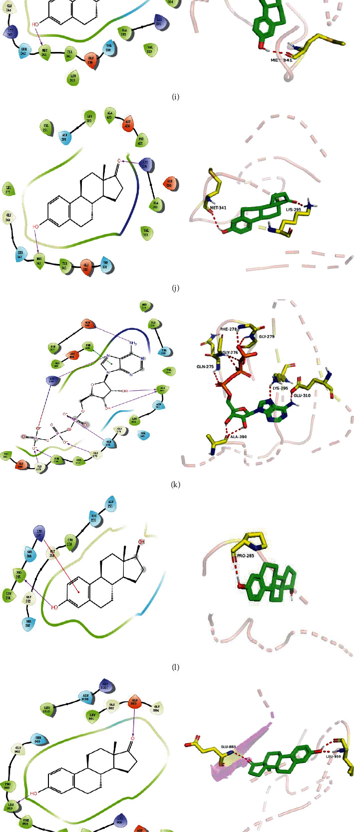
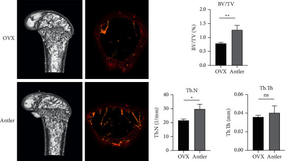
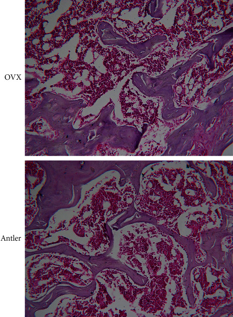
Deer velvet antlers are the young horns of male deer that are not ossified and densely overgrown. Velvet antler and its preparations have been widely used in the treatment of postmenopausal osteoporosis (PMOP) in recent years, although its mechanism of action in the human body remains unclear. To screen the effective ingredients and targets of velvet antler in the treatment of PMOP using network pharmacology and to explore the potential mechanisms of velvet antler action in such treatments, we screened the active ingredients and targets of velvet antler in the BATMAN-TCM database. We also screened the relevant targets of PMOP in the GeneCards and OMIM databases and then compared the targets at the intersection of both velvet antler and PMOP. We used Cytoscape 3.7.2 software to construct a network diagram of "disease-drug-components-targets" and a protein-protein interaction (PPI) network through the STRING database and screened out the core targets; the R language was then used to analyze the shared targets between antler and PMOP for GO-enrichment analysis and KEGG pathway-annotation analysis. Furthermore, we used the professional software Maestro 11.1 to verify the predictive analysis based on network pharmacology. Hematoxylin-eosin (H&E) staining and micro-CT were used to observe the changes in trabecular bone tissue, further confirming the results of network pharmacological analysis. The potentially effective components of velvet antler principally include 17β-E2, adenosine triphosphate, and oestrone. These components act on key target genes such as AKT1, IL6, MAPK3, TP53, EGFR, SRC, and TNF and regulate the PI3K/Akt-signaling and MAPK-signaling pathways. These molecules participate in a series of processes such as cellular differentiation, apoptosis, metabolism, and inflammation and can ultimately be used to treat PMOP; they reflect the overall regulation, network regulation, and protein interactions.
Copyright © 2021 Kuiting Guo et al.
Conflict of interest statement
The authors declare that they have no conflicts of interest.
Figures


Similar articles
-
Exploring the pharmacological components and effective mechanism of Mori Folium against periodontitis using network pharmacology and molecular docking.Arch Oral Biol. 2022 Jul;139:105391. doi: 10.1016/j.archoralbio.2022.105391. Epub 2022 Mar 21. Arch Oral Biol. 2022. PMID: 35430443
-
Potential Molecular Mechanisms of Ephedra Herb in the Treatment of Nephrotic Syndrome Based on Network Pharmacology and Molecular Docking.Biomed Res Int. 2022 Jul 5;2022:9214589. doi: 10.1155/2022/9214589. eCollection 2022. Biomed Res Int. 2022. PMID: 35837376 Free PMC article.
-
[Mechanism of active components of "Notoginseng Radix et Rhizoma-Drynariae Rhizoma" in treatment of osteoporosis based on network pharmacology and in vitro cell experiment].Zhongguo Zhong Yao Za Zhi. 2023 Feb;48(4):1087-1097. doi: 10.19540/j.cnki.cjcmm.20220926.501. Zhongguo Zhong Yao Za Zhi. 2023. PMID: 36872279 Chinese.
-
Well-known polypeptides of deer antler velvet with key actives: modern pharmacological advances.Naunyn Schmiedebergs Arch Pharmacol. 2024 Jan;397(1):15-31. doi: 10.1007/s00210-023-02642-y. Epub 2023 Aug 9. Naunyn Schmiedebergs Arch Pharmacol. 2024. PMID: 37555852 Review.
-
Exploration of the mechanism of Zisheng Shenqi decoction against gout arthritis using network pharmacology.Comput Biol Chem. 2021 Feb;90:107358. doi: 10.1016/j.compbiolchem.2020.107358. Epub 2020 Aug 8. Comput Biol Chem. 2021. PMID: 33243703 Review.
Cited by
-
Exploring the effect and mechanism of action of Jinlida granules (JLD) in the treatment of diabetes-associated cognitive impairment based on network pharmacology with experimental validation.Ann Med. 2025 Dec;57(1):2445181. doi: 10.1080/07853890.2024.2445181. Epub 2024 Dec 26. Ann Med. 2025. PMID: 39723533 Free PMC article.
-
Exploration of the Active Component and Mechanisms of Shengyu Decoction Against Myelosuppression Using Network Pharmacology and in vitro Experimental Validation.Drug Des Devel Ther. 2024 Jun 20;18:2405-2420. doi: 10.2147/DDDT.S458953. eCollection 2024. Drug Des Devel Ther. 2024. PMID: 38915868 Free PMC article.
-
DNA barcoding for the identification and authentication of medicinal deer (Cervus sp.) products in China.PLoS One. 2024 Jan 19;19(1):e0297164. doi: 10.1371/journal.pone.0297164. eCollection 2024. PLoS One. 2024. PMID: 38241246 Free PMC article.
-
Characterizing therapeutic effects of velvet antler using different omics strategies.J Tradit Complement Med. 2024 Aug 13;15(3):229-236. doi: 10.1016/j.jtcme.2024.08.002. eCollection 2025 May. J Tradit Complement Med. 2024. PMID: 40486283 Free PMC article. Review.
-
Bone metabolism associated with annual antler regeneration: a deer insight into osteoporosis reversal.Biol Direct. 2024 Nov 26;19(1):123. doi: 10.1186/s13062-024-00561-3. Biol Direct. 2024. PMID: 39593152 Free PMC article. Review.
References
-
- Li G., Li C., Li X., Liang W. The mechanism of estrogen regulating the imbalance of bone resorption-bone formation coupling in postmenopausal osteoporosis. Chinese medicine orthopedics . 2016;28(2):36–40.
-
- Somjen D., Katzburg S., Sharon O., Grafi-Cohen M., Knoll E., Stern N. The effects of estrogen receptors alpha- and beta-specific agonists and antagonists on cell proliferation and energy metabolism in human bone cell line. Journal of Cellular Biochemistry . 2011;112(2):625–632. doi: 10.1002/jcb.22959. - DOI - PubMed
-
- Silva I., Branco J. C. Denosumab: recent update in postmenopausal osteoporosis. Acta Reumatologica Portuguesa . 2012;37(4):302–313. - PubMed
-
- Asuman Dogan A., Nakipoglu-Yuzer G. F., Yldzgoren M. J., Ozgirgin N. Is age or the body mass index (BMI) more determinant of the bone mineral density (BMD)in geriatric women and men. Archives of Gerontology and Geriatrics . 2010;51(3):338–341. - PubMed
LinkOut - more resources
Full Text Sources
Research Materials
Miscellaneous
