. 2021 Oct 4:8:722446.
doi: 10.3389/fsurg.2021.722446. eCollection 2021.

The Effect of Waterpipe Tobacco Smoking on Bone Healing Following Femoral Fractures in Male Rats

Affiliations

The Effect of Waterpipe Tobacco Smoking on Bone Healing Following Femoral Fractures in Male Rats

Amirreza Sadeghifar et al. Front Surg. .

Abstract

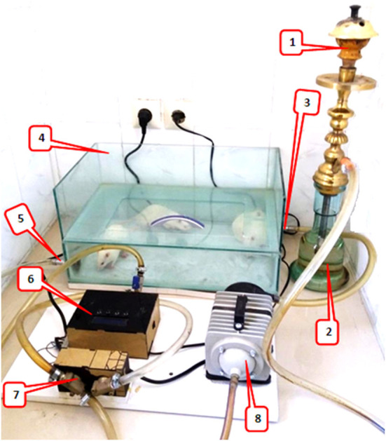
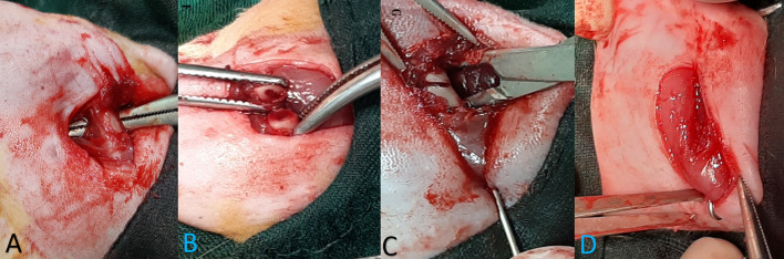
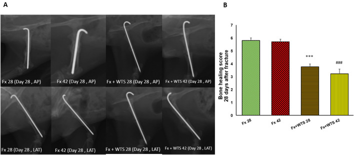
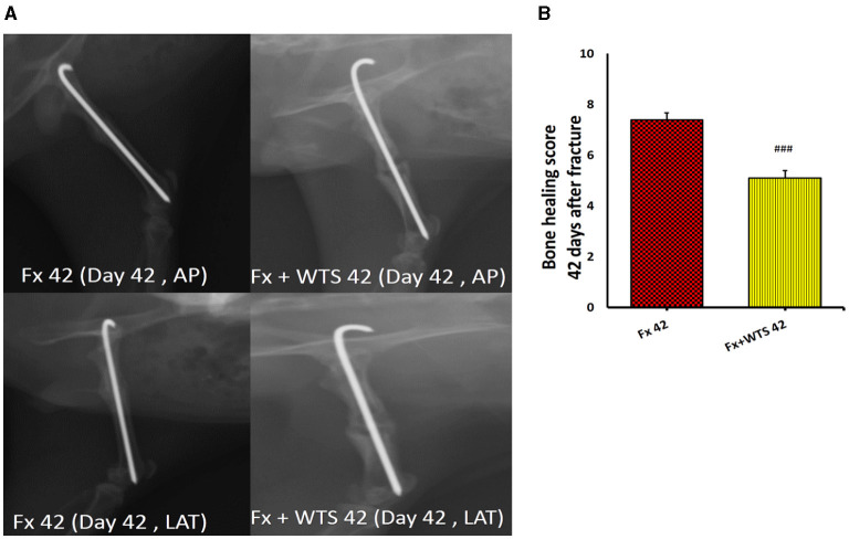
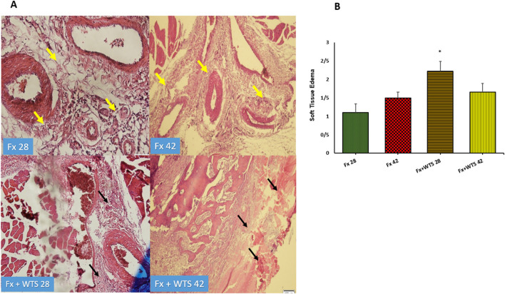
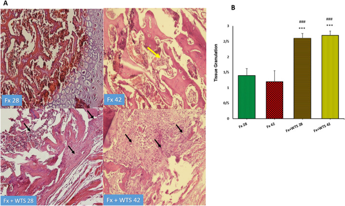
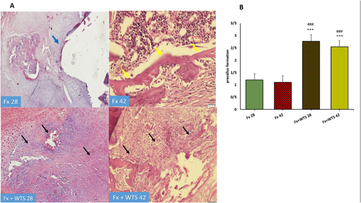
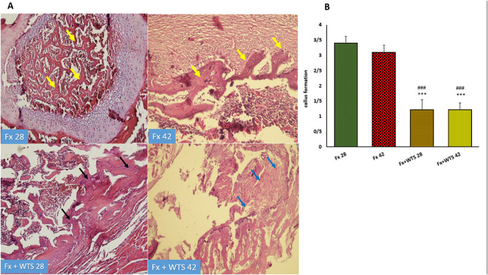
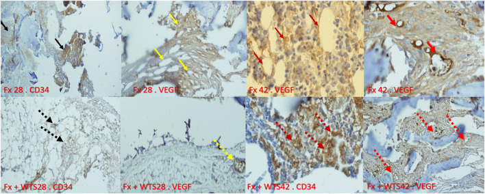
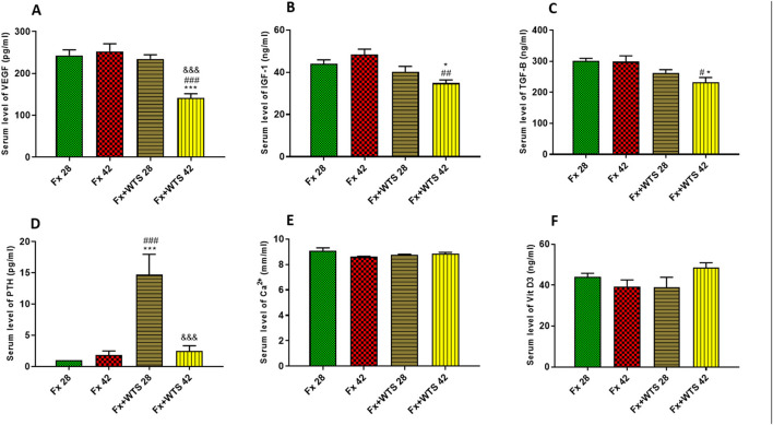
Background: Given the increasing use of waterpipe tobacco smoking in the world and its unknown effects on bone healing, this study investigated the repairing of femoral bone fractures in rats exposed to waterpipe tobacco smoking (WTS). Main Methods: This study involved 40 male Wistar rats that were divided into two groups, including the femoral fracture (Fx) and the Fx + WTS groups. Each group was divided into two subgroups that were evaluated for bone healing 28 and 42 days after femoral fracture. After fixing the fractured femur, the healing process was evaluated by radiography, pathological indicators, and a measurement of the blood levels of vascular endothelial growth factor (VEGF), parathyroid hormone (PTH), Ca ++, transforming growth factor-beta (TGF-β), and insulin-like growth factor 1 (IGF-1). Additionally, the density of VEGF and CD34 in fracture tissue was investigated by immunohistochemistry. Key Findings: Radiographic findings showed that factors related to the earlier stages of bone healing had higher scores in the Fx + WTS28 and 42 subgroups in comparison to the Fx groups. The density of VEGF and CD34 showed that the angiogenesis processes were different in the bone fracture area and callus tissue in the Fx +WTS subgroups. The serum levels of VEGF, TGF-β, and IGF-1 were significantly lower in the Fx +WTS42 group, and PTH in the Fx +WTS28 group was higher than that in the other groups. Significance: The findings showed the disturbance and delay in the femoral fracture union in rats exposed to hookah smoke. This is partly due to the reduction of molecular stimuli of bone synthesis and the attenuation of quantitative angiogenesis.

Keywords: TGF-β; angiogenesis markers; femur fracture; insulin growth factor-1; union; waterpipe tobacco smoking.


Conflict of interest statement

The authors declare that the research was conducted in the absence of any commercial or financial relationships that could be construed as a potential conflict of interest.

Figures

Figure 1
Figure 1
Waterpipe tobacco smoking setup: 1. the ceramic head of hookah for tobacco and charcoal, 2. water jar where the smoke passes, 3. fresh air/waterpipe smoke input, 4. a Plexiglass chamber (8 mm thick), placement for animals to inhale hookah smoke, 5. fresh air/waterpipe smoke output, 6. A timing controller for the sequence of operation of the pump and valve, 7. electronic valve, and 8. A vacuum pump that sucked tobacco smoke from the hookah and, with the help of a regulator, periodically controlled the blowing of smoke and fresh air through the valves into the chamber.
Figure 2
Figure 2
The stages of bone fracture. (A) Anesthesia induction was done by the injection of ketamine and xylazine and the maintenance of anesthesia during surgery with 1% isoflurane. (A) A lateral incision of the thigh was done and, through intermuscular plan femoral bone, has been exposed. (B) Osteotomy site. Osteotomy was performed with an orthopedic saw. Injected saline was used during the incision to prevent heat damage, and the cut was made completely transversely. (C) Retrogradely, the pin was inserted in the proximal part and exited from the greater trochanter; then, the reduction was established, and the pin was inserted and continued from the greater trochanter to the distal part of the femur. (D) Reduction was controlled visually and by radiography, then a pin was cut and buried under the muscles around the greater trochanter, and wound suturing was performed in anatomical layers.
Figure 3
Figure 3
Effects of waterpipe tobacco smoking (WTS) on serum cotinine levels in experimental groups. Fx 28: group subjected to room air for 28 days after surgery and a femoral bone fracture, Fx 42: group subjected to room air for 42 days after surgery and a femoral bone fracture, Fx + WTS 28: group subjected to WTS for 28 days after surgery and a femoral bone fracture, and Fx + WTS 42: group subjected to WTS for 42 days after surgery and a femoral bone fracture. The results are presented as mean ± SEM, n = 10. ***p < 0.001 vs. Fx 28; ###p < 0.001 vs. Fx 42; &p < 0.05 vs. Fx + WTS 28.
Figure 4
Figure 4
Radiographic evaluation revealed WTS decreased the process of fracture healing 10 days after fracture. (A) X-ray images of lateral and AP view of the femur. (B) The score of bone healing in experimental groups. Fx 28: group subjected to room air for 28 days after surgery and a femoral bone fracture, Fx 42: group subjected to room air for 42 days after surgery and a femoral bone fracture, Fx + WTS 28: group subjected to WTS for 28 days after surgery and a femoral bone fracture, Fx + WTS 42: group subjected to WTS for 42 days after surgery and a femoral bone fracture. The results are presented as mean ± SE, n = 10. **p < 0.01 vs. Fx 28; ##p < 0.01 vs. Fx 42.
Figure 5
Figure 5
Radiographic evaluation revealed WTS decreased the process of fracture healing in 28 days after fracture. (A) X-ray images of lateral and AP view of femur (B) the score of bone healing in experimental groups. Fx 28: group subjected to room air for 28 days after surgery and femoral bone fracture, Fx 42: group subjected to room air for 42 days after surgery and femoral bone fracture, Fx + WTS 28: group subjected to Waterpipe tobacco smoking for 28 days after surgery and femoral bone fracture, Fx+WTS 42: group subjected to Waterpipe tobacco smoking for 42 days after surgery and femoral bone fracture. The results are presented as mean ± SE, n = 10. ***p < 0.001 vs. Fx28; ###p < 0.001 vs. Fx 42.
Figure 6
Figure 6
Radiographic evaluation revealed WTS decreased the process of fracture healing 42 days after fracture. (A) X-ray images of lateral and AP view of the femur. (B) The score of bone healing in experimental groups. Fx 42: group subjected to room air for 42 days after surgery and a femoral bone fracture, Fx + WTS 42: group subjected to WTS for 42 days after surgery and a femoral bone fracture. The results are presented as mean ± SE, n = 10. ###p < 0.01 vs. Fx 42.
Figure 7
Figure 7
(A) Pathological representation of soft tissue edema. In the Fx 28 group, edema has decreased and indicates regular soft tissue without edema (the yellow arrows). In Fx 42 group, the same is true. In Fx + WTS 28 group, the soft tissue around the fracture is completely edematous and irregular and the beginning of the fibrosis process is observed (black arrows). In Fx + WTS 42 group, the surrounding tissue is completely fibrotic and edematous (black arrows). (B) The quantification of the soft tissue edema finding in experimental groups. Fx 28: group subjected to room air for 28 days after surgery and a femoral bone fracture, Fx 42: group subjected to room air for 42 days after surgery and a femoral bone fracture, Fx + WTS 28: group subjected to WTS for 28 days after surgery and a femoral bone fracture, Fx + WTS 42: group subjected to WTS for 42 days after surgery and a femoral bone fracture. The results are presented as mean ± SEM, n = 10. *p < 0.05 vs. Fx 28.
Figure 8
Figure 8
(A) Pathological representation of tissue granulation. In the Fx 28 group, tissue granulation is rare. In the Fx 42 group, the yellow arrow indicates that the tissue granulation has been removed and the bone marrow has been replaced. In the Fx + WTS 28 group, a tissue granulation fracture is observed, which is in the early stages of the healing process (black arrow). In the Fx + WTS 42 group, the black arrow indicates the fibrosis of the non-union, and tissue granulation has changed to fibrotic tissue (black arrow). (B) The quantification of the pathologic findings regarding tissue granulation in experimental groups. Fx 28: group subjected to room air for 28 days after surgery and a femoral bone fracture, Fx 42: group subjected to room air for 42 days after surgery and a femoral bone fracture, Fx + WTS 28: group subjected to WTS for 28 days after surgery and a femoral bone fracture, Fx + WTS 42: group subjected to WTS for 42 days after surgery and a femoral bone fracture. The results are presented as mean ± SEM, n = 10. ***p < 0.001 vs. Fx 28; ###p < 0.001 vs. Fx 42.
Figure 9
Figure 9
(A) Pathological representation of precallus formation. The Fx 28 group does not show precallus tissue, and the conversion of precallus to callus and regular bone is observed (blue arrow). In the Fx 42 group, bone callus formation and a regular osteoblastic rim forming callus can be seen (yellow arrows). The Fx + WTS 28 group indicates the beginning of the healing process and the beginning of bone and precallus tissue repair (black arrow). In the Fx + WTS 42 group, the precallus remnant and fibrotic non-union are observed (black arrow). (B) The quantification of pathologic findings regarding precallus formation in experimental groups. Fx 28: group subjected to room air for 28 days after surgery and a femoral bone fracture, Fx 42: group subjected to room air for 42 days after surgery and a femoral bone fracture, Fx + WTS 28: group subjected to WTS for 28 days after surgery and a femoral bone fracture, Fx + WTS 42: group subjected to WTS for 42 days after surgery and a femoral bone fracture. The results are presented as mean ± SEM, n = 10. ***p < 0.001 vs. Fx 28; ###p < 0.001 vs. Fx 42.
Figure 10
Figure 10
(A) Pathological representation of callus formation. In the Fx 28 group, fractures show bone blades that are properly formed and have regular bone marrow between them (yellow arrows). In the Fx 42 group, callus tissue is observed that has formed regularly (yellow arrows). In the Fx + WTS 28 group, pre-callus tissue is still observed, and the callus is irregularly formed and weakly shaped (black arrows). In the Fx + WTS 42 group, fibrotic non-union is observed (blue arrows). (B) The quantification of pathologic findings regarding callus formation in experimental groups. Fx 28: group subjected to room air for 28 days after surgery and a femoral bone fracture, Fx 42: group subjected to room air for 42 days after surgery and a femoral bone fracture, Fx + WTS 28: group subjected to WTS for 28 days after surgery and femoral bone fracture, Fx + WTS 42: group subjected to WTS for 42 days after surgery and femoral bone fracture. The results are presented as mean ± SEM, n = 10. ***p < 0.001 vs. Fx 28; ###p < 0.001 vs. Fx 42.
Figure 11
Figure 11
Immunohistochemical representation of the bone fracture area in experimental groups. Fx 28-CD34: The arteries that are properly formed around the fracture tissue (black arrow). Fx 28-vascular endothelial growth factor (VEGF): Represents open arteries and regular vascular tissue and is prominent around the fracture site (yellow arrow). Fx 42-VEGF: Indicates stained osteoblasts and clearly shows VEGF expression in osteoblasts that are active and regular (crimson arrow). Fx 42-VEGF: The vessels inside the callus are actively present and are supplying blood to the repair site (red arrow). Fx + WTS 28-CD34: Small, fragile vessels are shown around the repair site (black arrow point dots). Fx + WTS 28-VEGF: Small vessels and thrombosis are evident at the fracture site (yellow dashed arrow). Fx + WTS 42-CD34: Thrombotic and fragile arteries that are irregular and highly visible at the fracture site (crimson dashed arrow). Fx + WTS 42-VEGF: Small blood vessels inside the fibrotic tissue that are not functioning properly (red dashed arrow). Fx 28: group subjected to room air for 28 days after surgery and a femoral bone fracture, Fx 42: group subjected to room air for 42 days after surgery and a femoral bone fracture, Fx + WTS 28: group subjected to WTS for 28 days after surgery and a femoral bone fracture, Fx + WTS 42: group subjected to WTS for 42 days after surgery and a femoral bone fracture.
Figure 12
Figure 12
Effects of WTS on the serum levels of (A) VEGF, (B) insulin-like growth factor 1 (IGF-1), (C) transforming growth factor-beta (TGF-β), (D) parathyroid hormone (PTH), (E) Ca2+, and (F) vitamin D3 (Vit D3) in experimental groups. Fx 28: group subjected to room air for 28 days after surgery and a femoral bone fracture, Fx 42: group subjected to room air for 42 days after surgery and a femoral bone fracture, Fx + WTS 28: group subjected to WTS for 28 days after surgery and a femoral bone fracture, Fx + WTS 42: group subjected to WTS for 42 days after surgery and a femoral bone fracture. The results are presented as mean ± SEM, n = 7. *p < 0.05, ***p < 0.001 vs. Fx 28; #p < 0.05, ##p < 0.01, ###p < 0.001 vs. Fx 42; &&&p < 0.001 vs. Fx + WTS 28.

Similar articles

Cited by

References

    1. Badran M, Laher I. Waterpipe (shisha, hookah) smoking, oxidative stress and hidden disease potential. Redox Biol. (2020) 34:101455. 10.1016/j.redox.2020.101455 - DOI - PMC - PubMed
    1. Cobb CO, Shihadeh A, Weaver MF, Eissenberg T. Waterpipe tobacco smoking and cigarette smoking: a direct comparison of toxicant exposure and subjective effects. Nicotine Tob Res. (2011) 13:78–87. 10.1093/ntr/ntq212 - DOI - PMC - PubMed
    1. Hernigou J, Schuind F. Tobacco and bone fractures: A review of the facts and issues that every orthopaedic surgeon should know. Bone Jt Res. (2019) 8:255–65. 10.1302/2046-3758.86.BJR-2018-0344.R1 - DOI - PMC - PubMed
    1. Ma L, Zheng LW, Sham MH, Cheung LK. Uncoupled angiogenesis and osteogenesis in nicotine-compromised bone healing. J Bone Miner Res. (2010) 25:1305–13. 10.1002/jbmr.19 - DOI - PubMed
    1. Fischer C, Reiner C, Schmidmaier G, Doll J, Child C, Grützner PA, et al. . Safety study: Is there a pathologic IGF-1, PDGF and TGF-β cytokine expression caused by adjunct BMP-7 in tibial and femoral non-union therapy? Ther Clin Risk Manag. (2018) 14:691–7. 10.2147/TCRM.S160064 - DOI - PMC - PubMed