. 2021 Jul 5;12(9):1525-1539.
doi: 10.1039/d1md00124h. eCollection 2021 Sep 23.

Identification of a Zika NS2B epitope as a biomarker for severe clinical phenotypes

Affiliations

Identification of a Zika NS2B epitope as a biomarker for severe clinical phenotypes

Felix F Loeffler et al. RSC Med Chem. .

Abstract

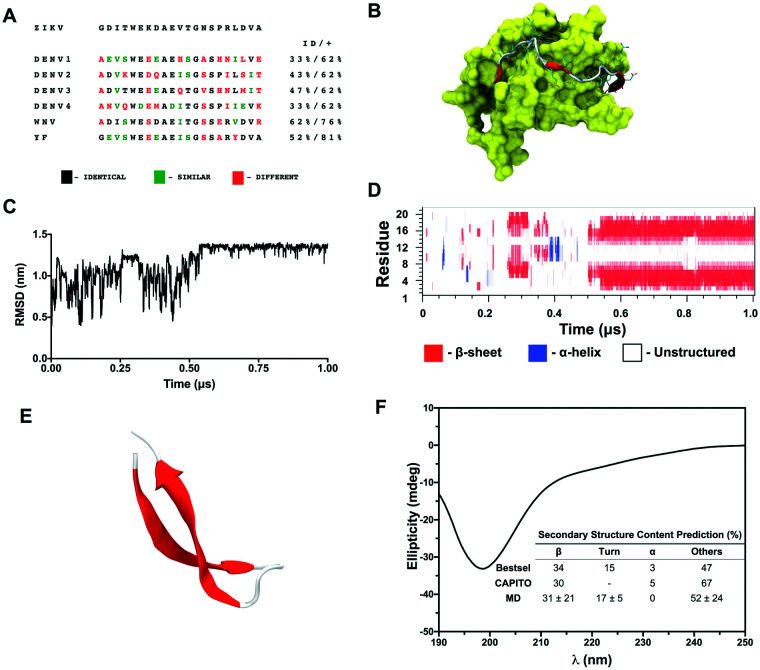
The identification of specific biomarkers for Zika infection and its clinical complications is fundamental to mitigate the infection spread, which has been associated with a broad range of neurological sequelae. We present the characterization of antibody responses in serum samples from individuals infected with Zika, presenting non-severe (classical) and severe (neurological disease) phenotypes, with high-density peptide arrays comprising the Zika NS1 and NS2B proteins. The data pinpoints one strongly IgG-targeted NS2B epitope in non-severe infections, which is absent in Zika patients, where infection progressed to the severe phenotype. This differential IgG profile between the studied groups was confirmed by multivariate data analysis. Molecular dynamics simulations and circular dichroism have shown that the peptide in solution presents itself in a sub-optimal conformation for antibody recognition, which led us to computationally engineer an artificial protein able to stabilize the NS2B epitope structure. The engineered protein was used to interrogate paired samples from mothers and their babies presenting Zika-associated microcephaly and confirmed the absence of NS2B IgG response in those samples. These findings suggest that the assessment of antibody responses to the herein identified NS2B epitope is a strong candidate biomarker for the diagnosis and prognosis of Zika-associated neurological disease.


Conflict of interest statement

A patent application for the epitope has been filed by TJ, NF, FFL, TM, and ETA. A patent application for the engineered protein carrying the identified NS2B epitope has been filed by RDL, DFC and IFTV. Otherwise, the authors have declared that they have no competing interests.

Figures

Fig. 1
Fig. 1. Sample classification used in the peptide array, resulting in the identification of the NS2B peptide. One hundred and twenty well-characterized serum samples collected from individuals aged 9 to 57 years old were divided into 8 groups, according to sample stratification through molecular and serological tests. Comparison groups included: 1. ZIKV+ acute samples with and without previous DENV infection (coloured in red); 2. ZIKV+ convalescent samples with and without previous DENV infection (coloured in blue); 3. ZIKV+ acute and convalescent samples with low anti-DENV antibody titers (coloured in magenta); 4. NeuroZIKV samples with and without previous DENV infection (coloured in green). GBS stands for Guillain–Barré syndrome.
Fig. 2
Fig. 2. Peptide array screening and identification of putative ZIKV NS2B peptides. (A) Peptide array screening showing the selection of 401 peptides covering the ZIKV NS1 and NS2B proteins. The IgM and IgG antibody profiles were determined for a control group (n = 8) and for a set of acute and convalescent serum samples from 103 individuals according to the following groups: i) acute Zika patients without dengue infection history (n = 14), ii) acute Zika patients with dengue infection history (n = 17), iii) acute Zika patients with low antibody titers against dengue (n = 3), iv) convalescent Zika patients without dengue infection history (n = 14), v) convalescent Zika patients with dengue infection history (n = 17), vi) convalescent Zika patients with low antibody titers against dengue (n = 3), vii) Zika infections with neurological manifestations (NeuroZIKV) without dengue infection history (n = 11), viii) NeuroZIKV infections with dengue infection history (n = 24). The IgM and IgG antibody response profiles are combined in the figure. (B) Identification of putative ZIKV NS2B epitopes. Immunoreactivity of the ZIKV NS2B peptides was individually assessed against the serum samples groups described in Fig. 2A. Bars represent the median fluorescence intensity for individual peptides. The four peptides showing the highest fluorescence signals in the array are highlighted in red.
Fig. 3
Fig. 3. Summary of the statistical data analysis. (A) Scores and (B) loadings plots of 2-component PCA model including control group, convalescent ZIKV+ (DENV−, DENV+ and DEN low) and NeuroZIKV, DENV− patients; and PLS-DA results for the 1-latent variable global model, including (C) ROC curve, (D) VIP scores plot for PLS-DA model and (E) predicted values of samples.
Fig. 4
Fig. 4. Summary of the additional PLS-DA models. (A) ROC curve and (B) VIP scores plot of PLS-DA to discriminate NeuroZIKV, DENV− from non-severe ZIKV patients (group VII vs. groups IV–VI); (C) ROC curve and (D) distribution classification histogram of patients on validation step for PLS-DA model to discriminate non-severe ZIKV, DENV− (group IV) from NeuroZIKV (groups VII, VIII) patients; (E) ROC curve and (F) VIP scores plot of PLS-DA to discriminate NeuroZIKV, DENV− from NeuroZIKV, DENV+ patients (group VII vs. group VIII).
Fig. 5
Fig. 5. Sequence and structural analyses of the identified NS2B peptide. (A) Sequence comparison of the 21-mer peptide based on the ZIKV NS2B epitope with the most frequently occurring strains of DENV, WNV, and YFV (residues are color-coded to highlight identity, chemical similarity, and differences. ‘ID’ stands for identity; a ‘+’ indicates similarity degree). (B) Structure representation of ZIKV NS2B bound to NS3 based on PDB ID 5H6V. The latter is shown as a yellow surface. NS2B is shown as cartoon and sticks, where β-strand regions are shown as red arrows, unstructured regions by a white tube and atoms in sticks color-coded as cyan for carbon, red for oxygen and blue for nitrogen. (C) Root-mean-square deviation of the NS2B-based peptide as a function of simulation time. The same region in PDB ID 5H6V was used as a reference. (D) Secondary structure content of the peptide as a function of simulation time. (E) Representative structure of the folded peptide in cartoon (β strands are shown in red and unstructured regions in white). (F) Far-UV circular dichroism (CD) spectra of the NS2B peptide in acetate buffer collected at 25 °C. Predicted secondary structure content based on experimental data is shown in the graph.
Fig. 6
Fig. 6. Structural characterization in silico of the engineered protein carrying the identified NS2B peptide. (A) Native structure of the scaffold protein (left) and structure of the NS2B-grafted protein after design/molecular dynamics (right), where the epitope region is shown in yellow and the native conformation of the epitope in NS2B is superimposed to it and displayed in cyan. The percentage indicates the sequence identity between the designed protein and the native scaffold. ΔΔG stands for the respective change of free energy of folding after the design protocol in Rosetta units (REU). (B) RMSD analysis for the backbone atoms for the native scaffold (black), for the NS2B-grafted designed protein (red) and comparison of the epitope with its native structure (blue). (C) Flexibility analysis using the RMSF values for the backbone atoms of the native scaffold (black) and NS2B-grafted (red). The region where the epitope was grafted is highlighted in yellow. (D) Solvent accessibility surface (SASA) indicating the exposure per residue of the epitope in its native structure (black) and in the designed protein (red). (E) Time-dependent secondary structure map indicating the maintenance of structural motifs during the MD simulation.
Fig. 7
Fig. 7. Detection of total IgG antibodies against the NS2B epitope engineered in the NS2B-grafted protein in paired samples from mothers and babies with ZIKV-associated microcephaly through ELISA. (A) Scatter plot representation of NS2B-specific IgG antibodies detection in serum samples from ZIKV naïve (ZIKV−/DENV− (n = 8) and ZIKV−/DENV+ (n = 8), negative controls) individuals, ZIKV+/DENV− individuals (n = 7, positive controls) and mothers who delivered babies with ZIKV-associated microcephaly and their progeny (n = 21 pairs). The results are shown as mean values over all measurements and corresponding standard error of the mean (S.E.M.). Dots represent individual measurements. A dashed line indicates the median of the positive control group for comparison purposes. The p-values of two-tailed Mann–Whitney test are indicated above each group comparison. (B) Comparison of the detection of anti-NS2B-IgG in mother–children paired samples. Each sample was tested in duplicate and the mean values are shown. A dashed line indicates the assay cut-off as calculated from a ROC curve (Fig. S6†). Gray lines connect paired samples. MC stands for ‘Microcephaly’.

References

    1. Faria N. R. Azevedo R. Kraemer M. U. G. Souza R. Cunha M. S. Hill S. C. Theze J. Bonsall M. B. Bowden T. A. Rissanen I. Rocco I. M. Nogueira J. S. Maeda A. Y. Vasami F. Macedo F. L. L. Suzuki A. Rodrigues S. G. Cruz A. C. R. Nunes B. T. Medeiros D. B. A. Rodrigues D. S. G. Queiroz A. L. N. da Silva E. V. P. Henriques D. F. da Rosa E. S. T. de Oliveira C. S. Martins L. C. Vasconcelos H. B. Casseb L. M. N. Simith D. B. Messina J. P. Abade L. Lourenco J. Alcantara L. C. J. de Lima M. M. Giovanetti M. Hay S. I. de Oliveira R. S. Lemos P. D. S. de Oliveira L. F. de Lima C. P. S. da Silva S. P. de Vasconcelos J. M. Franco L. Cardoso J. F. Vianez-Junior J. Mir D. Bello G. Delatorre E. Khan K. Creatore M. Coelho G. E. de Oliveira W. K. Tesh R. Pybus O. G. Nunes M. R. T. Vasconcelos P. F. C. Science. 2016;352:345. doi: 10.1126/science.aaf5036. - DOI - PMC - PubMed
    1. Brito C. Acta Med. Port. 2015;28:679. doi: 10.20344/amp.7341. - DOI - PubMed
    1. Schuler-Faccini L. Ribeiro E. M. Feitosa I. M. Horovitz D. D. Cavalcanti D. P. Pessoa A. Doriqui M. J. Neri J. I. Neto J. M. Wanderley H. Y. Cernach M. El-Husny A. S. Pone M. V. Serao C. L. Sanseverino M. T. Brazilian Medical Genetics Society-Zika Embryopathy Task F. Morb. Mortal. Wkly. Rep. 2016;65:59. doi: 10.15585/mmwr.mm6503e2. - DOI - PubMed
    1. Heymann D. L. Hodgson A. Sall A. A. Freedman D. O. Staples J. E. Althabe F. Baruah K. Mahmud G. Kandun N. Vasconcelos P. F. Bino S. Menon K. U. Lancet. 2016;387:719. doi: 10.1016/S0140-6736(16)00320-2. - DOI - PMC - PubMed
    1. WHO, Zika Causality Statement, https://www.who.int/emergencies/zika-virus/causality/en/, (accessed 26.03.2019, 2019)