. 2022 Mar;45(2):353-365.
doi: 10.1002/jimd.12450. Epub 2021 Nov 3.

The potential and limitations of intrahepatic cholangiocyte organoids to study inborn errors of metabolism

Affiliations

The potential and limitations of intrahepatic cholangiocyte organoids to study inborn errors of metabolism

Vivian Lehmann et al. J Inherit Metab Dis. 2022 Mar.

Abstract

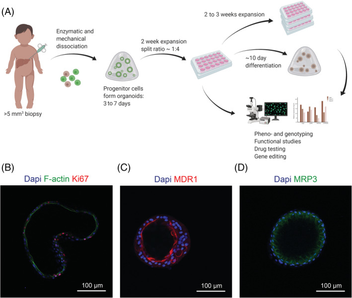
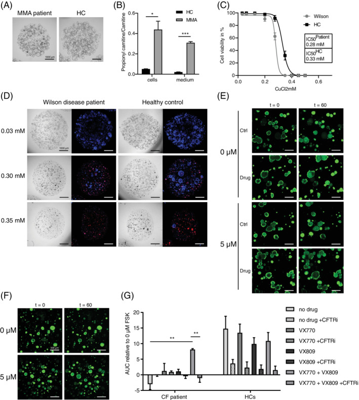
Inborn errors of metabolism (IEMs) comprise a diverse group of individually rare monogenic disorders that affect metabolic pathways. Mutations lead to enzymatic deficiency or dysfunction, which results in intermediate metabolite accumulation or deficit leading to disease phenotypes. Currently, treatment options for many IEMs are insufficient. Rarity of individual IEMs hampers therapy development and phenotypic and genetic heterogeneity suggest beneficial effects of personalized approaches. Recently, cultures of patient-own liver-derived intrahepatic cholangiocyte organoids (ICOs) have been established. Since most metabolic genes are expressed in the liver, patient-derived ICOs represent exciting possibilities for in vitro modeling and personalized drug testing for IEMs. However, the exact application range of ICOs remains unclear. To address this, we examined which metabolic pathways can be studied with ICOs and what the potential and limitations of patient-derived ICOs are to model metabolic functions. We present functional assays in patient ICOs with defects in branched-chain amino acid metabolism (methylmalonic acidemia), copper metabolism (Wilson disease), and transporter defects (cystic fibrosis). We discuss the broad range of functional assays that can be applied to ICOs, but also address the limitations of these patient-specific cell models. In doing so, we aim to guide the selection of the appropriate cell model for studies of a specific disease or metabolic process.

Keywords: Wilson disease; cystic fibrosis; inborn errors of metabolism; intrahepatic cholangiocyte organoids; methylmalonic acidemia; patient-specific in vitro modeling.


Conflict of interest statement

The authors declare no potential conflict of interest.

Figures

FIGURE 1
FIGURE 1
Overview of organoid generation, use and characteristics. (A) Organoid generation from biopsy to in vitro culture can be achieved within 3 weeks, whereafter pheno‐ and genotyping, functional assays, drug testing, gene editing, and/or differentiation can take place. (B) Organoids express proliferation marker Ki67 and apical marker F‐actin in expansion conditions (EM). After differentiation (DM), organoids condense and display more mature liver functions and stronger polarization, as exemplified by the apical transporter MDR1 and the basolateral transporter MRP3. Nuclear staining is shown with Dapi
FIGURE 2
FIGURE 2
Functional assays revealing disease phenotypes in patient ICOs. (A) Representative culture of MMA patient and HC ICOs under EM. (B) Branched‐chain amino acid metabolism defect exemplified by significantly increased propionylcarnitine to carnitine ratio in ICOs of an MMA patient (n = 2; *, P‐value = .0221 in cells; ***, P‐value = .0009 in medium). (C) Copper metabolism defect exemplified by increased sensitivity to copper (CuCl2) toxicity in ICOs of a Wilson disease patient compared to a HC after 72 hours of exposure (n = 12). (D) Brightfield and IF images of the viability assay in C. showing key concentrations of CuCl2. Red signal corresponds to necrosis marker propidium iodide; blue signal indicates DNA. (E‐G) Impaired apical transport can be studied in ICOs as exemplified by defect and rescued CFTR function (***, P‐value <.0001) in ICOs of a Cystic fibrosis patient. (E,F) Representative images of CF patient (E) and HC (F) ICO swelling. (G) AUC of CF patient (n = 3) and HC (n = 6) ICO swelling with or without the drugs VX809 and VX‐770 (**, P‐value <.0031) and with or without CFTR inhibitor (**, P‐value <.0028) at 5 μM FSK relative to 0 μM FSK. AUC, area under the curve; CF, cystic fibrosis; EM, expansion condition; FSK, forskolin; HC, healthy control
FIGURE 3
FIGURE 3
Visualizations of log2 fold changes in gene expression of IEM genes in expanded (EM, n = 2) and differentiated (DM, n = 2) ICOs, fibroblasts (Fibro, n = 2) and whole liver tissue (n = 2). log2 fold changes are relative to the mean expression of the genes across all fibroblast and ICO DM samples. IEM genes are divided into metabolic categories Amino Acid Metabolism (A), Pentose Phosphate Pathway (B), Carbohydrate Metabolism (other) (C), Citric acid cycle (D), Lysosomal Metabolism (E), Trace element and Metal Metabolism (F), Glycogen Metabolism (G), Oxidative Phosphorylation (H), Porphyrin and Haem Metabolism (I), Fatty Acid and Ketone Body Metabolism (J), Glycolysis and Glyconeogenesis (K), Peroxisomal Metabolism (L), Glycan Metabolism (M), Xenobiotics Metabolism (N), Lipid Metabolism (O), Metabolism of vitamins and (nonprotein) cofactors (P), Nucleotide Metabolism (Q), Other (R)
FIGURE 4
FIGURE 4
(A) Schematic representation of a hepatocyte showing metabolic functions and approximate organelle locations. Organelle sizes not to scale. (B) TEM images of HC ICO cells in EM and DM displaying differences in ICO wall thickness. Arrows indicate mucus fields. Red squares indicate magnification regions shown in C. (C) TEM images of HC ICO cells in EM and DM displaying the major organelles visible in these cells (left), mucus and villus presence (right), and peroxisomes (D). TEM, transmission electron microscopy; DM, differentiation condition; EM, expansion condition; Gol, golgi; Gly, glycogen rosettes; Lys, lysosomes; Mit, mitochondria; Nuc, nucleus; Per, peroxisomes; ER, endoplasmic reticulum; Vil, villi

References

    1. Ferreira CR, van Karnebeek CDM, Vockley J, Blau N. A proposed nosology of inborn errors of metabolism. Genet Med. 2019;21(1):102‐106. doi:10.1038/s41436-018-0022-8 - DOI - PMC - PubMed
    1. Waters D, Adeloye D, Woolham D, Wastnedge E, Patel S, Rudan I. Global birth prevalence and mortality from inborn errors of metabolism: a systematic analysis of the evidence. J Glob Health. 2018;8(2). doi:10.7189/jogh.08.021102 - DOI - PMC - PubMed
    1. Stenton SL, Prokisch H. Genetics of mitochondrial diseases: identifying mutations to help diagnosis. EBioMed. 2020;56:102784. doi:10.1016/j.ebiom.2020.102784 - DOI - PMC - PubMed
    1. Tarasenko TN, Mcguire PJ. The liver is a metabolic and immunologic organ: a reconsideration of metabolic decompensation due to infection in inborn errors of metabolism (IEM). Mol Genet Metab. 2017;121:283‐288. doi:10.1016/j.ymgme.2017.06.010 - DOI - PMC - PubMed
    1. Sato T, Vries RG, Snippert HJ, et al. Single Lgr5 stem cells build crypt‐villus structures in vitro without a mesenchymal niche. Nat. 2009;459(7244):262‐265. doi:10.1038/NATURE07935 - DOI - PubMed

Publication types

MeSH terms

Substances