

In Vitro Models Mimicking Immune Response in the Skin
- PMID: 34672130
- PMCID: PMC8542468
- DOI: 10.3349/ymj.2021.62.11.969
In Vitro Models Mimicking Immune Response in the Skin
Abstract
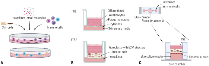
The skin is the first line of defense of our body, and it is composed of the epidermis and dermis with diverse immune cells. Various in vitro models have been investigated to recapitulate the immunological functions of the skin and to model inflammatory skin diseases. The simplest model is a two-dimensional (2D) co-culture system, which helps understand the direct and indirect cell-to-cell interactions between immune and structural cells; however, it has limitations when observing three-dimensional (3D) interactions or reproducing skin barriers. Conversely, 3D skin constructs can mimic the human skin characteristics in terms of epidermal and dermal structures, barrier functions, cell migration, and cell-to-cell interaction in the 3D space. Recently, as the importance of neuro-immune-cutaneous interactions in the inflammatory response is emerging, 3D skin constructs containing both immune cells and neurons are being developed. A microfluidic culture device called "skin-on-a-chip," which simulates the structures and functions of the human skin with perfusion, was also developed to mimic immune cell migration through the vascular system. This review summarizes the in vitro skin models with immune components, focusing on two highly prevalent chronic inflammatory skin diseases: atopic dermatitis and psoriasis. The development of these models will be valuable in studying the pathophysiology of skin diseases and evaluating the efficacy and toxicity of new drugs.
Keywords: Atopic dermatitis; immune system; in vitro techniques; lab-on-a-chip devices; psoriasis; skin equivalent.
© Copyright: Yonsei University College of Medicine 2021.
Conflict of interest statement
The authors have no potential conflicts of interest to disclose.
Figures
References
-
- Reus AA, Usta M, Krul CA. The use of ex vivo human skin tissue for genotoxicity testing. Toxicol Appl Pharmacol. 2012;261:154–163. - PubMed
-
- Ackermann K, Borgia SL, Korting HC, Mewes KR, Schäfer-Korting M. The Phenion Full-Thickness Skin model for percutaneous absorption testing. Skin Pharmacol Physiol. 2010;23:105–112. - PubMed
Publication types
MeSH terms
Grants and funding
LinkOut - more resources
Full Text Sources
Other Literature Sources
Medical
Miscellaneous
