. 2022 Dec 29;118(16):3250-3268.
doi: 10.1093/cvr/cvab321.

Interferon-stimulated gene 15 pathway is a novel mediator of endothelial dysfunction and aneurysms development in angiotensin II infused mice through increased oxidative stress

Affiliations

Interferon-stimulated gene 15 pathway is a novel mediator of endothelial dysfunction and aneurysms development in angiotensin II infused mice through increased oxidative stress

María González-Amor et al. Cardiovasc Res. .

Abstract

Aims: Interferon-stimulated gene 15 (ISG15) encodes a ubiquitin-like protein that induces a reversible post-translational modification (ISGylation) and can also be secreted as a free form. ISG15 plays an essential role as host-defence response to microbial infection; however, its contribution to vascular damage associated with hypertension is unknown.

Methods and results: Bioinformatics identified ISG15 as a mediator of hypertension-associated vascular damage. ISG15 expression positively correlated with systolic and diastolic blood pressure and carotid intima-media thickness in human peripheral blood mononuclear cells. Consistently, Isg15 expression was enhanced in aorta from hypertension models and in angiotensin II (AngII)-treated vascular cells and macrophages. Proteomics revealed differential expression of proteins implicated in cardiovascular function, extracellular matrix and remodelling, and vascular redox state in aorta from AngII-infused ISG15-/- mice. Moreover, ISG15-/- mice were protected against AngII-induced hypertension, vascular stiffness, elastin remodelling, endothelial dysfunction, and expression of inflammatory and oxidative stress markers. Conversely, mice with excessive ISGylation (USP18C61A) show enhanced AngII-induced hypertension, vascular fibrosis, inflammation and reactive oxygen species (ROS) generation along with elastin breaks, aortic dilation, and rupture. Accordingly, human and murine abdominal aortic aneurysms showed augmented ISG15 expression. Mechanistically, ISG15 induces vascular ROS production, while antioxidant treatment prevented ISG15-induced endothelial dysfunction and vascular remodelling.

Conclusion: ISG15 is a novel mediator of vascular damage in hypertension through oxidative stress and inflammation.

Keywords: Endothelial dysfunction; ISG15; Inflammation; Oxidative stress; Vascular remodelling.


Conflict of interest statement

Conflict of interest: none declared.

Figures

Graphical Abstract
Graphical Abstract
Figure 1
Figure 1
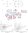
ISG15 is a potential mediator of vascular damage. Interactions between UBA7 (A) or USP18 (B) with other proteins involved in hypertension, obtained with Ingenuity Pathways Analysis software. Protein–protein interaction network between hypertension-related proteins and ISG15 by STRING/Cytoscape (C). Correlation between ISG15 mRNA and systolic blood pressure (D) or carotid intima-media thickness (E) in human peripheral blood mononuclear cells of asymptomatic patients. AU, arbitrary units.
Figure 2
Figure 2
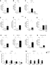
Angiotensin II (AngII) induces ISG15 expression at the vascular level and in macrophages. (A) Isg15 mRNA levels in vascular smooth muscle cells (VSMC) from C57BL/6J mice incubated or not with IFN-γ (50 U/mL, 2 h). (B) Ifng mRNA levels in aorta from mice untreated (control) and treated with AngII (1.44 mg/kg/day, 2 weeks). (C) Isg15 mRNA levels in aorta from C57BL/6J mice incubated or not with AngII (1 μmol/L 6 h) in the absence or presence of anti-IFN-γ antibody (5 μg/mL). Isg15 mRNA levels (D) and secreted ISG15 protein (E) in aorta from mice untreated (Control) and treated with AngII. (F) Isg15 mRNA levels in aorta from 6-month-old normotensive (Wistar Kyoto, WKY) and spontaneously hypertensive (SHR) rats. (G) Isg15 mRNA levels in aorta from mice untreated (Control) and treated with AngII or AngII and hydralazine plus hydrochlorothiazide (HH, hydralazine: 20 mg/kg/day; hydrochlorothiazide: 6 mg/kg/day i.p). ISG15 mRNA levels in VSMC (H), human microvascular endothelial cells (HMEC-1) (I), and human aortic endothelial cells (HAEC) (J) incubated with AngII (1 nmol/L, 4 h). Isg15 mRNA levels in peritoneal macrophages from untreated mice (Control) and treated with AngII (K). mRNA levels of ISGylation enzymes (UBA7, Ube2L6, and HERC5/Herc6) and de-ISGylation enzyme USP18 in aorta from Control and AngII-infused mice (L) and HMEC-1 treated or not with AngII (1 nmol/L, 4 h) (M). *P < 0.05 by Student’s t-test or one-way ANOVA.
Figure 3
Figure 3
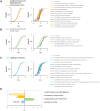
Quantitative proteomics demonstrates that ISG15 promotes a coordinated alteration of cardiovascular remodelling, extracellular matrix and cardiovascular function categories. Aortic tissue samples from WT and ISG15−/− treated or not with Angiotensin II (AngII) were subjected to quantitative proteomics using multiplexed isobaric labelling and LC–MS/MS. The quantitative data were analysed using the SBT model to detect coordinated protein changes in functional categories. The distributions of quantitative protein values (Zq) are plotted for four clusters (A) cardiovascular remodelling and extracellular matrix, (B) cardiovascular function and (C) immune system. Panels on the left display the cumulative distribution of Zq from proteins belonging to each cluster. Panels on the right display the protein values belonging to the related GO terms which compose each cluster. Protein values (Zq) are log2 fold changes in AngII-treated ISG15−/− compared to AngII-treated WT, normalized with respect to untreated samples, expressed in units of standard deviation. (D) Standardized log2 fold changes (Zc) of the four category clusters (*FDR < 0.05; **FDR < 0.01). The complete set of proteins belonging to each cluster is listed in Supplementary material online, Table S11.
Figure 4
Figure 4
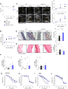
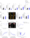
ISG15 participates in Angiotensin II (AngII)-induced hypertension and vascular damage. (A) Systolic blood pressure (SBP) measured by tail-cuff plethysmography (WT and WT AngII: n = 12, ISG15−/−: n = 10, ISG15−/− AngII: n = 11); *P < 0.05 vs. untreated, #P < 0.05 vs. WT AngII by two-way ANOVA. (B) Aortic Masson stain and media thickness quantification; *P < 0.05 by one-way ANOVA. Structural (C and D) and mechanical parameters (E) in small mesenteric arteries (SMA) (n = 7–12); *P < 0.05 vs. untreated by two-way ANOVA or *P < 0.05 by one-way ANOVA. (F) Quantification of internal elastic lamina structure of SMA; *P < 0.05 by one-way ANOVA. Concentration-response curves to acetylcholine (ACh; G and I) and diethylamine NONOate (DEA-NO; H and J) of aorta or SMA (n = 7–12); *P < 0.05 vs. untreated, #P < 0.05 vs. WT AngII by two-way ANOVA.
Figure 5
Figure 5
ISGylation participates in angiotensin II (AngII)-induced hypertension and vascular damage. (A) Survival curve of AngII-treated WT and USP18C61A mice (n = 15–27); #P < 0.05 vs. WT AngII by log-rank (Mantel–Cox) test. (B) Systolic blood pressure (SBP) measured by tail-cuff plethysmography (WT and WT AngII: n = 9, USP18C61A and USP18C61A AngII: n = 11); *P < 0.05 vs. untreated, #P < 0.05 vs. WT AngII by two-way ANOVA. (C) Representative ultrasound images and quantification of maximal diameter in ascending aorta (AsAo) or abdominal aorta (AbAo) (WT and WT AngII: n = 12, USP18C61A: n = 8, USP18C61A AngII: n = 9); *P < 0.05 vs. untreated, #P < 0.05 vs. WT AngII by two-way ANOVA. (D) Representative aortic elastic Van Gieson (EVG) staining and quantification; *P < 0.05 by one-way ANOVA. (E) Representative aortic Sirius red staining and quantification; *P < 0.05 by one-way ANOVA. (F) Aortic mRNA expression of fibrotic markers; *P < 0.05 by one-way ANOVA. (GJ) Concentration-response curves to acetylcholine (ACh) and diethylamine NONOate (DEA-NO) of aorta and SMA (n = 6–9); *P < 0.05 vs. untreated by two-way ANOVA.
Figure 6
Figure 6
ISG15 participates in angiotensin II (AngII)-induced inflammation and oxidative stress. (A) Quantitative proteomics analysis of proteins related to the vascular redox state cluster. Results were presented as in Figure 3. The functional category abundance change value for this cluster was Zc = −1.83 (log2 fold change in ISG15−/− AngII with respect to WT AngII, normalized with respect to untreated samples, in units of standard deviation), which is statistically significant at FDR <0.05. The complete set of proteins belonging to this cluster is listed in Supplementary material online, Table S11. (B) Quantitative redox proteomics shows increased abundance of peptides containing reversibly oxidized Cys sites in AngII-treated WT mice in comparison with ISG15−/− mice. Shown are the cumulative distributions of Zp, the standardized log2 ratio of oxidized-Cys-containing peptides in WT AngII, ISG15−/− AngII and ISG15−/−, with respect to WT. The graph also shows the distribution of Zp for all the peptides of the experiment (in grey). The oxidized-Cys-containing peptides of the WT AngII sample is significantly increased with respect to the WT sample (P < 0.002, Kolmogorov–Smirnov test). (C) Representative dihydroethidium (DHE) fluorescence and quantification in media and adventitia layers. (D) Aortic mRNA expression of Ifng, Ptgs2, Ccl2, Adgre1, and Cd3e. *P < 0.05 by one-way ANOVA.
Figure 7
Figure 7
ISGylation increases inflammation and reactive oxygen species generation that participate of vascular damage. (A) Aortic mRNA expression of Ifng, Ptgs2, Ccl2, Adgre1, and Cd3e; *P < 0.05 by one-way ANOVA. (B) Aortic mRNA expression of Cybb; *P < 0.05 by one-way ANOVA. (C) Representative dihydroethidium (DHE) fluorescence and quantification in media and adventitia layers. *P < 0.05 by one-way ANOVA. Because experiments were run simultaneously, data from WT and WT-angiotensin II (AngII) from (A) and (C) are the same as Figure 6D and C, respectively. Representative images for DHE staining are different. (D) Survival curve of AngII-infused USP18C61A mice treated or not with the antioxidant tempol (0.288 nmol/kg/day) (n = 10–11); P < 0.05 vs. USP18C61A AngII by log-rank (Mantel–Cox) test. (E) Representative images of aorta from USP18C61A-AngII mice treated or not with tempol (F) systolic blood pressure (SBP) measured by tail-cuff plethysmography (n = 8–12); P < 0.05 vs. USP18C61A AngII by two-way ANOVA. Structural (G and H) and mechanical (I) parameters in small mesenteric arteries (n = 4–8); P < 0.05 vs. USP18C61A AngII by two-way ANOVA.

References

    1. Brandes RP. Endothelial dysfunction and hypertension. Hypertension 2014;64:924–928. - PubMed
    1. Laurent S, Boutouyrie P.. The structural factor of hypertension: large and small artery alterations. Circ Res 2015;116:1007–1021. - PubMed
    1. Sun X-N, Li C, Liu Y, Du L-J, Zeng M-R, Zheng X-J, Zhang W-C, Liu Y, Zhu M, Kong D, Zhou L, Lu L, Shen Z-X, Yi Y, Du L, Qin M, Liu X, Hua Z, Sun S, Yin H, Zhou B, Yu Y, Zhang Z, Duan S-Z.. T-cell mineralocorticoid receptor controls blood pressure by regulating interferon-gamma. Circ Res 2017;120:1584–1597. - PubMed
    1. Norlander AE, Madhur MS, Harrison DG.. The immunology of hypertension [published correction appears in J Exp Med. 2018 Jan 5]. J Exp Med 2018;215:21–33. - PMC - PubMed
    1. Caillon A, Paradis P, Schiffrin EL.. Role of immune cells in hypertension. Br J Pharmacol 2019;176:1818–1828. - PMC - PubMed

Publication types

MeSH terms