Hypoxia promotes the proliferation of mouse liver sinusoidal endothelial cells: miRNA-mRNA expression analysis
- PMID: 34672871
- PMCID: PMC8806994
- DOI: 10.1080/21655979.2021.1988371
Hypoxia promotes the proliferation of mouse liver sinusoidal endothelial cells: miRNA-mRNA expression analysis
Abstract
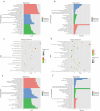
During the initial stage of liver regeneration (LR), hepatocytes and liver sinusoidal endothelial cells (LSECs) initiate regeneration in a hypoxic environment. However, the role of LSECs in liver regeneration in hypoxic environments and their specific molecular mechanism is unknown. Therefore, this study aimed to explore the miRNA-mRNA network that regulates the proliferation of LSECs during hypoxia. In this study, first, we found that the proliferation ability of primary LSECs treated with hypoxia was enhanced compared with the control group, and then whole transcriptome sequencing was performed to screen 1837 differentially expressed (DE) genes and 17 DE miRNAs. Subsequently, the bioinformatics method was used to predict the target genes of miRNAs, and 309 pairs of interacting miRNA-mRNA pairs were obtained. Furthermore, the miRNA-gene action network was established using the negative interacting miRNA-mRNA pairs. The selected mRNAs were analyzed by Gene Ontology (GO) enrichment analysis and Kyoto Encyclopedia of Genes and Genomes (KEGG) analysis, and biological processes (BP) and signal pathways related to LSEC proliferation that were significantly enriched in GO-BP and KEGG were selected. Finally, 22 DE genes and 17 DE miRNAs were screened and the network was created. We also successfully verified the significant changes in the top six genes and miRNAs using qRT-PCR, and the results were consistent with the sequencing results. This study proposed that a specific miRNA-mRNA network is associated with hypoxia-induced proliferation of LSECs, which will assist in elucidating the potential mechanisms involved in hypoxia-promoting liver regeneration during LR.
Keywords: Liver sinusoidal endothelial cells; bioinformatics; hypoxia; liver regeneration; microrna.
Conflict of interest statement
No potential conflict of interest was reported by the author(s).
Figures


Similar articles
-
Hypoxia maintains the fenestration of liver sinusoidal endothelial cells and promotes their proliferation through the SENP1/HIF-1α/VEGF signaling axis.Biochem Biophys Res Commun. 2021 Feb 12;540:42-50. doi: 10.1016/j.bbrc.2020.12.104. Epub 2021 Jan 12. Biochem Biophys Res Commun. 2021. PMID: 33445109
-
Identification of Serum Exosome-Derived circRNA-miRNA-TF-mRNA Regulatory Network in Postmenopausal Osteoporosis Using Bioinformatics Analysis and Validation in Peripheral Blood-Derived Mononuclear Cells.Front Endocrinol (Lausanne). 2022 Jun 9;13:899503. doi: 10.3389/fendo.2022.899503. eCollection 2022. Front Endocrinol (Lausanne). 2022. PMID: 35757392 Free PMC article.
-
Comprehensive analysis of lncRNA-miRNA-mRNA networks during osteogenic differentiation of bone marrow mesenchymal stem cells.BMC Genomics. 2022 Jun 7;23(1):425. doi: 10.1186/s12864-022-08646-x. BMC Genomics. 2022. PMID: 35672672 Free PMC article.
-
Integrated miRNA-mRNA network revealing the key molecular characteristics of ossification of the posterior longitudinal ligament.Medicine (Baltimore). 2020 May 22;99(21):e20268. doi: 10.1097/MD.0000000000020268. Medicine (Baltimore). 2020. PMID: 32481304 Free PMC article.
-
The role of microRNAs in the different phases of liver regeneration.Expert Rev Gastroenterol Hepatol. 2023 Jul-Dec;17(10):959-973. doi: 10.1080/17474124.2023.2267422. Epub 2023 Oct 24. Expert Rev Gastroenterol Hepatol. 2023. PMID: 37811642 Review.
Cited by
-
SENP1 attenuates hypoxia‑reoxygenation injury in liver sinusoid endothelial cells by relying on the HIF‑1α signaling pathway.Mol Med Rep. 2024 Apr;29(4):64. doi: 10.3892/mmr.2024.13188. Epub 2024 Mar 1. Mol Med Rep. 2024. PMID: 38426545 Free PMC article.
-
MiR-424-5p suppresses tumor growth and progression by directly targeting CHEK1 and activating cell cycle pathway in Hepatocellular Carcinoma.Heliyon. 2024 Sep 11;10(18):e37769. doi: 10.1016/j.heliyon.2024.e37769. eCollection 2024 Sep 30. Heliyon. 2024. PMID: 39309825 Free PMC article.
-
Screening and Biological Function Analysis of miRNA and mRNA Related to Lung Adenocarcinoma Based on Bioinformatics Technology.J Oncol. 2022 Aug 31;2022:4339391. doi: 10.1155/2022/4339391. eCollection 2022. J Oncol. 2022. PMID: 36090902 Free PMC article.
-
The long noncoding RNA noncoding RNA activated by DNA damage (NORAD)-microRNA-496-Interleukin-33 axis affects carcinoma-associated fibroblasts-mediated gastric cancer development.Bioengineered. 2021 Dec;12(2):11738-11755. doi: 10.1080/21655979.2021.2009412. Bioengineered. 2021. PMID: 34895039 Free PMC article.
References
-
- Fausto N, Campbell JS, Riehle KJ.. Liver regeneration. Hepatology. 2006;43(S1):S45–53. - PubMed
-
- Valizadeh A, Majidinia M, Samadi-Kafil H, et al. The roles of signaling pathways in liver repair and regeneration. J Cell Physiol. 2019;234(9):14966–14974. - PubMed
-
- Fraser R, Dobbs BR, Rogers GW.. Lipoproteins and the liver sieve: the role of the fenestrated sinusoidal endothelium in lipoprotein metabolism, atherosclerosis, and cirrhosis. Hepatology. 1995;21:863–874. - PubMed
Publication types
MeSH terms
Substances
LinkOut - more resources
Full Text Sources
Other Literature Sources
