. 2021 Nov;297(5):101312.
doi: 10.1016/j.jbc.2021.101312. Epub 2021 Oct 19.

A single amino acid mutation in the mouse MEIG1 protein disrupts a cargo transport system necessary for sperm formation

Affiliations

A single amino acid mutation in the mouse MEIG1 protein disrupts a cargo transport system necessary for sperm formation

Wei Li et al. J Biol Chem. 2021 Nov.

Abstract

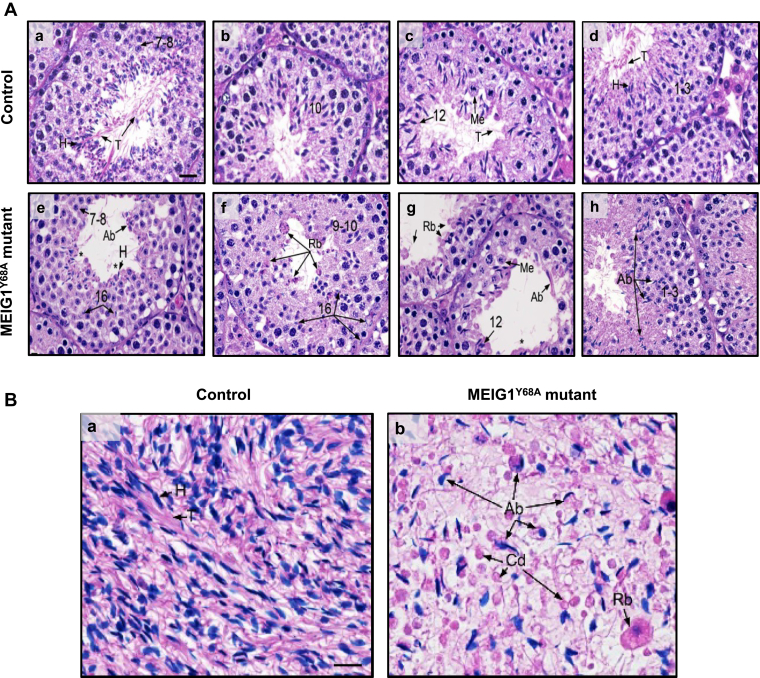
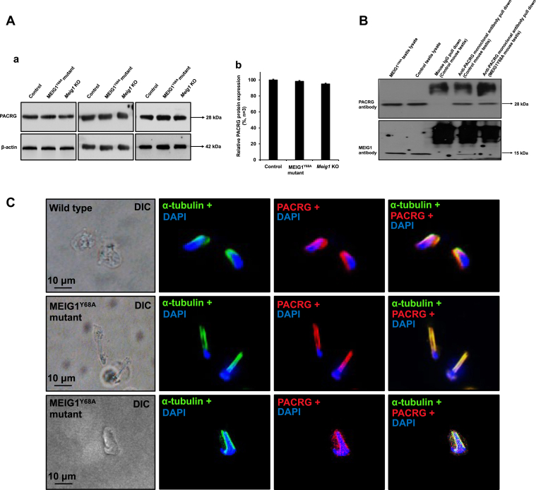
Mammalian spermatogenesis is a highly coordinated process that requires cooperation between specific proteins to coordinate diverse biological functions. For example, mouse Parkin coregulated gene (PACRG) recruits meiosis-expressed gene 1 (MEIG1) to the manchette during normal spermiogenesis. Here we mutated Y68 of MEIG1 using the CRISPR/cas9 system and examined the biological and physiological consequences in mice. All homozygous mutant males examined were completely infertile, and sperm count was dramatically reduced. The few developed sperm were immotile and displayed multiple abnormalities. Histological staining showed impaired spermiogenesis in these mutant mice. Immunofluorescent staining further revealed that this mutant MEIG1 was still present in the cell body of spermatocytes, but also that more MEIG1 accumulated in the acrosome region of round spermatids. The mutant MEIG1 and a cargo protein of the MEIG1/PACRG complex, sperm-associated antigen 16L (SPAG16L), were no longer found to be present in the manchette; however, localization of the PACRG component was not changed in the mutants. These findings demonstrate that Y68 of MEIG1 is a key amino acid required for PACRG to recruit MEIG1 to the manchette to transport cargo proteins during sperm flagella formation. Given that MEIG1 and PACRG are conserved in humans, small molecules that block MEIG1/PACRG interaction are likely ideal targets for the development of male contraconception drugs.

Keywords: MEIG1; PACRG; SPAG16; cargo transport; male fertility; manchette; single amino acid mutation; spermiogenesis.


Conflict of interest statement

Conflict of interest The authors declare that they have no conflicts of interest with the contents of this article.

Figures

Figure 1
Figure 1
Generation of MEIG1Y68Amutant mouse model.A, examination of testicular MEIG1 expression in the indicated mice. (a) Representative Western blot results to examine testicular MEIG1 protein expression in the control, Y68A homozygous mutant, and global Meig1 knockout mice. MEIG1 is missing in the global knockout mice, but is still present in the Y68A mutant mice. (b) Quantification of the relative MEIG1 protein expression normalized by β-actin. Error bars represent the standard deviation (n = 3). (∗), p < 0.05. B, mislocalization of Y68A mutant MEIG1 in round and elongating spermatids. The testicular cells were double stained with MEIG1 antibody (red) and the acrosome marker lectin PNA (green), or a manchette marker α-tubulin (green). Wild-type MEIG1 is localized in cell bodies of spermatocytes (panel a, arrow) and round spermatids (panel b, dashed arrows), and migrates to the manchette of elongating spermatids (panel c, arrow head). Y68A mutant MEIG1 is still present in cell bodies of spermatocytes (panel d, f, arrows), but is highly concentrated in the acrosome of round spermatids (panel e, dashed arrows); it is not present in the manchette of the remaining elongating spermatids (panel f, arrow heads).
Figure 2
Figure 2
Y68 single amino acid defect in MEIG1 causes male infertility associated with abnormal sperm morphology, significantly reduced sperm number and motility.A, fertility of control and MEIG1Y68A mutant mice. Ten controls and ten MEIG1Y68A mutant mice were examined. Fertility and litter size were recorded for each mating. Notice that all mutant females had normal fertility; however, all mutant males were infertile. B, normal testis/body weight of control and Y68A mutant mice. C, morphological examination of epididymal sperm from the control (left) and mutant (right) mice by light microscopy at low magnification. Notice that sperm density of the control mice is higher than those observed in the MEIG1Y68A mice under the same dilution. Sperm in the control mice showed normal morphology, but short tails (lower, left inset), vesicles in the flagella (lower, right inset), and different thicknesses along the tails (upper, inset) were frequently observed in the mutant mice, Bar = 10 μm. D, sperm number was significantly reduced in the mutant mice. E, percentage of abnormal sperm of control and the mutant mice. F, percentage of motile sperm of control and the mutant mice. “n” represents the number of mice analyzed. Data are expressed as Mean ± SD. Statistically significant differences (∗), p < 0.05.
Figure 3
Figure 3
Spermiogenesis defect in the MEIG1Y68Amutant mice.A, testis histology of control (a–d) and homozygous Y68A mutant mice (e–h). (a) Stage VII–VIII showing normal spermatogenesis with step 7 to 8 round spermatids. Step 16 spermatids are aligned by their heads (H) at the lumen, with extensive tails (T). Bar = 25 μm (for all photos). (b) Stage X showing normal step 10 spermatids just beginning to show elongation of the nucleus. (c) Stage XII showing normal meiotic division of spermatocytes (Me) and step 12 elongating spermatids, with their long tails (T) extending into the lumen. (d) Stage I–III showing normal step 1 to 3 round spermatids and elongating spermatids with heads (H) aligned and tails (T) extending into the lumen. (e) Mutant stage VII–VIII showing normal step 7 to 8 round spermatids, but abnormal elongating spermatids (Ab). The abnormal elongating spermatid heads (H) are surrounded by cytoplasm and appear rounded without an extension of the tails (∗). The heads of step 16 spermatids are seen deep within the epithelium, where they have been phagocytized by the Sertoli cells. (f) Mutant stage IX–X showing disorganization of the step 9 to 10 spermatids, with possibly some abnormal shapes. Failure of spermiation is present with rounded cytoplasm of residual bodies (Rb) containing the heads of sperm without tails. Some individual step 16 spermatid heads are also seen having been phagocytized by Sertoli cells. (g) Mutant stage XII showing normal meiotic division (Me) of spermatocytes, but disorganization of the step 12 elongating spermatids. The elongating spermatids are abnormal, lacking tails in many cases (∗) and showing abnormal tails (Ab) with excessive cytoplasm in other cases. (h) Mutant stage I–III showing normal step 1 to 3 round spermatids, but abnormal heads of elongating spermatids (Ab) and few tails extending into the lumen. B, the control mouse cauda epididymis showing highly concentrated normal sperm in the lumen with heads (H) and tails (T) aligned (a). The MEIG1Y68A mutant cauda epididymis (b) showing very low concentration of sperm, with high incidence of sperm abnormalities (Ab), including absent tails, excess cytoplasm, and short tails. Round bodies (Rb) in the lumen appear to be larger residual bodies and smaller cytoplasmic droplets (Cd). Few sperm tails are present. Bar = 20 mm (for all photos).
Figure 4
Figure 4
TEM images of seminiferous tubules of a control mouse and MEIG1Y68Amutant mice. A number of normally developed sperm were released to the lumen of the seminiferous tubules of a control mouse. Normal chromatin condensation (dotted arrow), well-condensed head (arrow), and normally developed flagella can be observed. The arrow heads point to the middle piece (right, inset) and the principle piece (left, inset) in (a). (b) shows few sperm present in the lumen in the MEIG1Y68A mutant mice. Abnormal “9 + 2” axoneme structure surrounded by abnormally accessory structures (arrow heads in b–e) were frequently observed. Increased number of the lysosomes were seen in the degrading abnormal flagellum (stars, b). Multiple elongating spermatids in the seminiferous tubule were wrapped in 1 cell membrane, indicating phagocytized by Sertoli cells (arrowheads, e). Abnormal condensed head (triangles, f) and chromatin can be seen in the mutant mice (arrows, in g and h).
Figure 5
Figure 5
The localization and expression of PACRG were not changed in the MEIG1Y68Amutant mice.A, examination of testicular PACRG expression in the indicated mice by Western blot analysis. (a) Representative Western blot results to examine testicular PACRG protein expression in the control and Y68A homozygous mutant mice; (b) Quantification of the relative PACRG protein expression normalized by β-actin. Error bars represent the standard deviation (n = 3). There was no significant difference in PACRG level between the control and the MEIG1Y68A mutant mice. B, PACRG binds to wild-type MEIG1 but not the Y68A mutant MEIG1 in the testis. Co-IP experiment was conducted using testicular lysates from the control and Y68A mutant mice. The PACRG antibody pulled down wild-type MEIG1 but not the mutant MEIG1. C, localization of PACRG in germ cells of MEIG1Y68A mutant mice. PACRG is present in the manchette of elongating spermatids in wild-type mice (upper panel). In MEIG1Y68A mutant mice, the localization of PACRG was not changed (middle and bottom panels).
Figure 6
Figure 6
MEIG1Y68Amutation changes localization of SPAG16L in elongating spermatids.A, examination of testicular SPAG16L expression in the indicated mice by Western blot analysis. (a) Representative Western blot results to examine testicular SPAG16L protein expression in the control and Y68A homozygous mutant mice; (b) Quantification of the relative SPAG16L protein expression normalized by β-actin. Error bars represent the standard deviation (n = 3). There was no significant difference in SPAG16L level between the control and the MEIG1Y68A mutant mice. B, localization of SPAG16L in germ cells of wild-type and MEIG1Y68A mutant mice. In control mice, SPAG16L staining (red) was observed in the cytoplasm of spermatocytes (panel a, b, arrows) and round spermatids (panel a, dotted arrows), and the manchette of elongating spermatids (panel b, arrow heads), as evaluated by double staining with an anti-α-tubulin antibody (green). In MEIG1Y68A mutant mice, SPAG16L is still present in cytoplasm of spermatocytes (panel c, arrow) and round spermatids (panel c, dotted arrows); however, it is not present in the manchette of elongating spermatids (panel d, arrow head).
Figure 7
Figure 7
Working model of MEIG1/PACRG complex in transporting cargo in the manchette. MEIG1 is recruited to the manchette by PACRG. SPAG16L, a sperm structure protein localized in the central apparatus of axoneme is a cargo of MEIG1/PACRG complex. Disruption of MEIG1/PACRG complex, such as mutation of Y68 on the MEIG1 protein shuts down the transport system.

References

    1. Don J., Wolgemuth D.J. Identification and characterization of the regulated pattern of expression of a novel mouse gene, meg1, during the meiotic cell cycle. Cell Growth Differ. 1992;3:495–505. - PubMed
    1. Zhang Z., Shen X., Gude D.R., Wilkinson B.M., Justice M.J., Flickinger C.J., Herr J.C., Eddy E.M., Strauss J.F. MEIG1 is essential for spermiogenesis in mice. Proc. Natl. Acad. Sci. U. S. A. 2009;106:17055–17060. - PMC - PubMed
    1. Salzberg Y., Eldar T., Karminsky O.D., Itach S.B., Pietrokovski S., Don J. Meig1 deficiency causes a severe defect in mouse spermatogenesis. Dev. Biol. 2010;338:158–167. - PubMed
    1. Don J., Winer M.A., Wolgemuth D.J. Developmentally regulated expression during gametogenesis of the murine gene meg1 suggests a role in meiosis. Mol. Reprod. Dev. 1994;38:16–23. - PubMed
    1. Chen-Moses A., Malkov M., Shalom S., Ever L., Don J. A switch in the phosphorylation state of the dimeric form of the Meg1 protein correlates with progression through meiosis in the mouse. Cell Growth Differ. 1997;8:711–719. - PubMed

Publication types

MeSH terms