

doi: 10.1016/j.resmer.2021.100862.
Epub 2021 Oct 2.
Two-photon microscopy analysis reveals different pulmonary damage after infection by influenza or SARS-CoV-2
Affiliations
- PMID: 34673456
- PMCID: PMC8486684
- DOI: 10.1016/j.resmer.2021.100862
Item in Clipboard
Two-photon microscopy analysis reveals different pulmonary damage after infection by influenza or SARS-CoV-2
Respir Med Res.
2021 Nov.
No abstract available
Keywords: 2-photon microscopy; COVID-19; Influenza.
Conflict of interest statement
Declaration of Competing Interest None. The images have not been previously published.
Figures
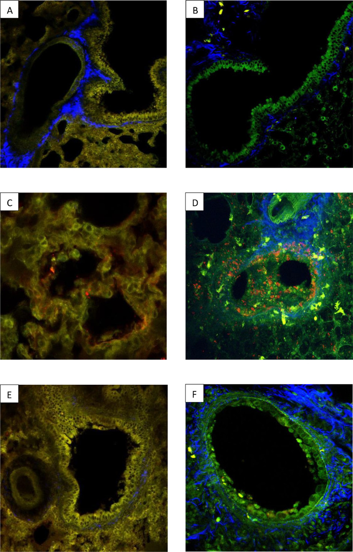
Comparative analysis with 2MP of lung injury after infection with SARS-Cov2 or Influenzavirus. The images above show two 2-photon micrographs (2PM) of normal hamster (A) and mouse (B) lung parenchyma. Alveolar/bronchial epithelium tissue is shown in yellow (A) or green (B) and connective tissue (in both micrographs) is blue. Two 2PM micrographs of pulmonary parenchyma showing the distribution of SARS-Cov-2 (C) or the A/PR8 influenza (D) virus 72 h after infection. In (C) the hamster tissue was stained with the SARS-Cov-2 Spike antibody and a secondary antibody coupled with AF568, an orange-red dye. In (D) the mouse tissue was stained with an endogen fluorescence NS1 coupled with a red fluorescent marker (RFP). The images show that SARS-CoV-2 was localized exclusively on the alveolar epithelium whereas the influenza virus was predominantly localized the on bronchial and alveolar epithelia. Two 2PM micrographs of pulmonary bronchial epithelia 72 h after infection with either SARS-Cov-2 (E) or A/PR8 influenza (F) virus. In (E) the hamster tissue was stained with the SARS-Cov-2 Spike antibody and a secondary antibody coupled with AF568, an orange-red dye. In (F) the mouse tissue was stained with an endogen fluorescence NS1 coupled with a red fluorescent marker (RFP) to visualise the influenza virus. The bronchial epithelium (E) had a normal structure 72 h following infection with Sars-Cov-2 in contrast to the epithelium (F) which showed signs of severe injury following infection with Influenzavirus.
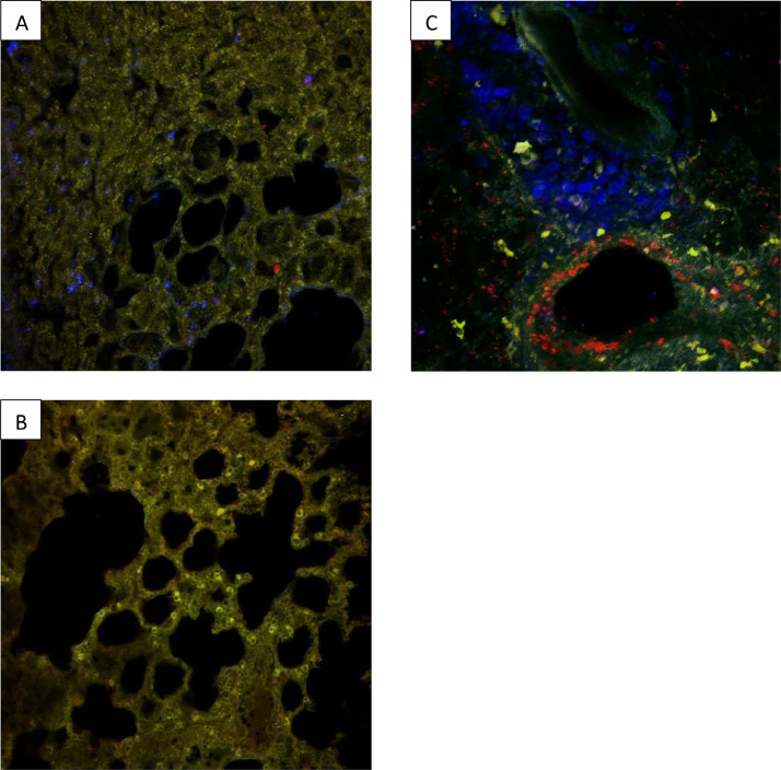
Comparison of three 2PM images showing localized macrophage recruitment in pulmonary epithelial tissue 72 h after infection with either SARS-Cov-2 virus (A) and (B), or A/PR8 influenza (C). In all slides an F4/80 antibody coupled with a blue BV421 fluorescent marker was used to stain the macrophages. In slides (A) and (B) the SARS-Cov-2 Spike antibody and a secondary antibody coupled with AF568 (an orange-red dye) were used to stain the SARS-Cov-2 virus and in slide (C) an endogenous, fluorescent NS1 coupled with a red fluorescent marker (RFP) were used to visualise the influenza virus. Macrophage recruitment in SARS-Cov-2 infected lungs was heterogenous, with diseased (A) and healthy areas (B) in the same organ. This variation in macrophage distribution differed from the general, diffuse macrophage recruitment from the perivascular area observed in lung tissue after infection with influenza (C).
References
Publication types
MeSH terms
LinkOut - more resources
Full Text Sources
Medical
Miscellaneous
