

Bioprosthetic Aortic Valve Degeneration: a Review from a Basic Science Perspective
- PMID: 34673516
- PMCID: PMC9054148
- DOI: 10.21470/1678-9741-2020-0635
Bioprosthetic Aortic Valve Degeneration: a Review from a Basic Science Perspective
Abstract
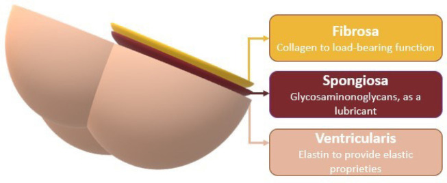
Introduction: The increase in the prevalence of aortic stenosis due to an aging population has led to an increasing number of surgical aortic valve replacements. Over the past 20 years, there has been a major shift in preference from mechanical to bioprosthetic valves. However, despite efforts, there is still no "ideal" bioprosthesis. It is crucial to understand the structure, biology, and function of native heart valves to design more intelligent, strong, durable, and physiological heart valve tissues.
Methods: A comprehensive review of the literature was performed to identify articles reporting the basic mechanisms of bioprosthetic valve dysfunction and the biology of native valve cells. Searches were run in PubMed, MEDLINE® (the Medical Literature Analysis and Retrieval System Online), and Google Scholar. Terms for subject heading and keywords search included "biological heart valve dysfunction", "bioprosthesis dysfunction", "bioprosthesis degeneration", and "tissue heart valves".
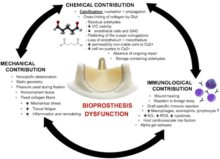
Results: All the relevant findings are summarized in the appropriate subsections. Structural dysfunction is a logical and expected consequence of the chemical, mechanical, and immunological processes that occur during fixation, manufacture, and implantation.
Conclusion: Biological prosthesis valve dysfunction is a clinically significant process. It has become a major issue considering the growing rate of bioprosthesis implantation and improved long-term patient survival. Understanding bioprosthetic aortic valve degeneration from a basic science perspective is a key point to improve technologic advances and specifications that lead to a new generation of bioprostheses.
Keywords: Aging; Aortic Valve; Aortic Valve Stenosis; Bioprosthesis; Immunology; Inflammation.
Conflict of interest statement
No conflict of interest.
Figures
References
COMPLEMENTARY REFERENCE
-
- Hilbert SL, Barrick MK, Ferrans VJ. Porcine aortic valve bioprostheses: A morphologic comparison of the effects of fixation pressure. J Biomed Mater Res. 1990;24(6):773–87. - PubMed
-
- Sacks MS, Smith DB, Hiester ED. The aortic valve microstructure: Effects of transvalvular pressure. J Biomed Mater Res. 1998;41(1):131–41. - PubMed
-
- Lo D, Vesely I. Biaxial strain distributions in expanded porcine bioprosthetic valves. J Heart Valve Dis. 2002;11(5):688–95. - PubMed
-
- Vesely I. Reconstruction of loads in the fibrosa and ventricularis of porcine aortic valves. ASAIO J. 1996;42(5) - PubMed
-
- Scott M, Vesely I. Morphology of porcine aortic valve cusp elastin. J Hear Valve Dis. 1996;5(5):464–71. - PubMed
Publication types
MeSH terms
LinkOut - more resources
Full Text Sources
Miscellaneous