. 2021 Oct 21;19(10):e3001434.
doi: 10.1371/journal.pbio.3001434. eCollection 2021 Oct.

The Plasmodium NOT1-G paralogue is an essential regulator of sexual stage maturation and parasite transmission

Affiliations

The Plasmodium NOT1-G paralogue is an essential regulator of sexual stage maturation and parasite transmission

Kevin J Hart et al. PLoS Biol. .

Abstract

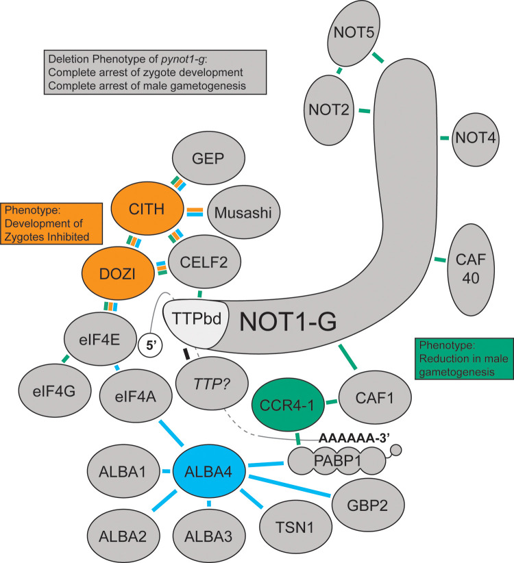
Productive transmission of malaria parasites hinges upon the execution of key transcriptional and posttranscriptional regulatory events. While much is now known about how specific transcription factors activate or repress sexual commitment programs, far less is known about the production of a preferred mRNA homeostasis following commitment and through the host-to-vector transmission event. Here, we show that in Plasmodium parasites, the NOT1 scaffold protein of the CAF1/CCR4/Not complex is duplicated, and one paralogue is dedicated for essential transmission functions. Moreover, this NOT1-G paralogue is central to the sex-specific functions previously associated with its interacting partners, as deletion of not1-g in Plasmodium yoelii leads to a comparable or complete arrest phenotype for both male and female parasites. We show that, consistent with its role in other eukaryotes, PyNOT1-G localizes to cytosolic puncta throughout much of the Plasmodium life cycle. PyNOT1-G is essential to both the complete maturation of male gametes and to the continued development of the fertilized zygote originating from female parasites. Comparative transcriptomics of wild-type and pynot1-g- parasites shows that loss of PyNOT1-G leads to transcript dysregulation preceding and during gametocytogenesis and shows that PyNOT1-G acts to preserve mRNAs that are critical to sexual and early mosquito stage development. Finally, we demonstrate that the tristetraprolin (TTP)-binding domain, which acts as the typical organization platform for RNA decay (TTP) and RNA preservation (ELAV/HuR) factors is dispensable for PyNOT1-G's essential blood stage functions but impacts host-to-vector transmission. Together, we conclude that a NOT1-G paralogue in Plasmodium fulfills the complex transmission requirements of both male and female parasites.


Conflict of interest statement

The authors have declared that no competing interests exist.

Figures

Fig 1
Fig 1. NOT1 is duplicated in the Aconoidasida class of apicomplexans.
Shown here are examples of NOT1 proteins from the Aconoidasida class of apicomplexans with a representative model eukaryote NOT1 protein from Saccharomyces cerevisiae. Protein and domain sizes are drawn to scale. Bioinformatically predicted domains were identified through BlastP searches and amino acid alignments. TTP, tristetraprolin.
Fig 2
Fig 2. PyNOT1 and PyNOT1-G have similar expression patterns and localize to cytosolic puncta.
Representative IFA images of (A) PyNOT1::GFP and (B) PyNOT1-G::GFP blood stage parasites show localization patterns typical for the CAF1/CCR4/NOT complex in eukaryotes. All samples were stained with DAPI and mouse anti-GFP to image PyNOT1::GFP or PyNOT1-G::GFP. Asexual blood stage parasites were counterstained with anti-PyACP and DAPI to identify ring, trophozoite, and schizont stages. Gametocytes were counterstained with anti-alpha tubulin (male) or anti-PyCITH (female). Scale bar is 5 μm. DIC, differential interference contrast; IFA, immunofluorescence assay.
Fig 3
Fig 3. PyNOT1-G is essential to the formation of male gametes.
(A) The blood stage growth of Py17XNL wild-type parasites and 2 independent clones of pynot1-g parasites was compared over the entire course of infection in biological triplicate, with each replicate done in technical triplicate. Maximal parasitemia of pynot1-g parasites was lower and resulted in slightly faster clearance by the mouse. (B) The number of gametocytes produced by Py17XNL WT-GFP parasites and 2 independent clones of pynot1-g parasites was measured by flow cytometry using the presence of GFP fluorescence in biological duplicate, with each replicate done in technical triplicate. Average values for each biological replicate, their median, and range are provided. Statistical comparisons used an unpaired t test with Welch correction. (C) Male gametogenesis was measured by DIC microscopy to count the number of exflagellation centers (“centers of movement”) per microscopic field using a 10× eyepiece and 40× objective lens. The same mice and time points were used as in panel A and were assessed in biological triplicate, with each replicate done in technical triplicate. A cross symbol (A, C) indicates that a single mouse infected with Py17XNL wild-type parasites was euthanized on day 15 due to parasitemia more than 10% as required by our approved vertebrate animal protocols. Error bars indicate the standard deviation. Underlying data are provided in S1 Data. DIC, differential interference contrast.
Fig 4
Fig 4. PyNOT1-G is essential to the development of zygotes resulting from female gametocytes.
(A) A genetic cross experiment with Py17XNL wild-type parasites (colorless) and pynot1-g (GFP+) transgenic parasites was conducted to assess effects upon parasite transmission. Mice were infected with a total of 10,000 mixed blood stage parasites consisting of only wild-type parasites, an equal mixture of wild-type and transgenic parasites, or only transgenic parasites. Mosquito transmission occurred on the day where peak exflagellation was observed for mice infected with only wild-type parasites, and colorless and GFP+ oocysts were counted 7 days post-blood meal by fluorescence and DIC microscopy. An illustration of the experimental readout/interpretation (top) and the results (bottom) are provided. Experiments were conducted in biological triplicate, with error bars denoting SEM. (B) A genetic cross experiment with Py17XNL WT-mScarlet, WT-GFP, and pynot1-g (GFP+) transgenic parasites was conducted using in vitro ookinete culture conditions. Equal volumes of infected blood at comparable parasitemias were mixed as indicated and were scored by live fluorescence microscopy for the presence/absence of mScarlet and GFP signals. Representative images of the WT-mScarlet x WT-GFP genetic cross are provided to show zygote, retort, and ookinete stages (top). Results of all tested genetic crosses are provided (bottom) from experiments conducted in biological duplicate. Parasites resulting from the WT-mScarlet x pynot1-g (GFP+) cross could not develop beyond the zygote stage. Scale bars are 5 μm (zygote, retort) or 10 μm (ookinete). Underlying data are provided in S1 Data. DIC, differential interference contrast.
Fig 5
Fig 5. Extensive dysregulation of mRNAs in pynot1-g schizonts and gametocytes.
Comparative RNA-seq of wild-type or pynot1-g transgenic parasites was conducted using mixed male and female gametocytes or schizonts that were enriched through a subtractive magnetic depletion approach. (A) VST-transformed raw counts were used as input values to generate a PCA plot to demonstrate sample clustering. (B) Heatmaps of the expression of 50 selected transcripts for gametocytes (left) and schizonts (right) further illustrate clustering and the effect of deleting pynot1-g. (C and D) MA plots generated from the estimated shrunken log2 fold changes are shown for gametocytes (C) and schizonts (D), with data points shaded in red indicating p < 0.05 (comparing pynot1-g/WT). Data points that do not meet this threshold are denoted as open triangles (oriented up or down). (E and F) Plotted are the −log10 FDR vs the log2 fold change values for differentially expressed transcripts as per DEseq2, with transcripts shaded based upon their fold change values (comparing pynot1-g/WT). Similar gametocyte- and early mosquito stage-enriched transcripts were significantly lower in abundance in both stages. (G and H) CirGO plots of GO terms for gametocytes (left) and schizonts (right) are provided that highlight key biological processes that are impacted by PyNOT1-G. FDR, false discovery rate; GO, gene ontology; PCA, principal component analysis; VST, variance-stabilizing transformation.
Fig 6
Fig 6. The putative TTP-binding domain is dispensable for all essential blood stage functions of PyNOT1-G.
Clonal transgenic parasites overexpressing the predicted TTP-binding domain fused to GFP (TTPbd::GFP) from the safe harbor p230p genomic locus were compared to Py17XNL WT-GFP parasites for (A) asexual blood stage growth and (B) male gametogenesis. Clonal transgenic parasites expressing a variant of PyNOT1-G where the N-terminal TTPbd has been replaced with GFP were compared to Py17XNL WT-GFP parasites for (C) asexual blood stage growth and (D) male gametogenesis. A cross symbol (A, B) indicates that a single mouse infected with TTPbd::GFP Clone 2 parasites was euthanized on day 15 due to parasitemia more than 10% as required by our approved vertebrate animal protocols. Error bars indicate the standard deviation. Underlying data are provided in S1 Data. TTP, tristetraprolin.
Fig 7
Fig 7. A proposed composite interaction network of NOT1-G.
Crosslinking IP datasets for PbDOZI and PbCITH (orange lines), PyCCR4-1 (green lines), and PyALBA4 (light blue lines) were used to propose a composite interaction network with PyNOT1-G. The placement of some proteins is based upon known direct interactions from the literature (e.g., NOT1-G, CAF1, CCR4, CAF40, NOT4, NOT2, NOT5, PABP1, CELF2, DOZI, eIF4F (eIF4E, eIF4A, eIF4G)), as are the placements of the 5′ m7G cap next to the cap-binding protein eIF4E and the poly(A) tail with PABP1. Deletion phenotypes for pynot1-g match or exceed those of pbdozi, pyccr4–1, and the pycaf1 truncation and lead to a complete arrest in male parasite development and in development of female parasites beyond the zygote stage. TTP, tristetraprolin.

Similar articles

Cited by

References

    1. World Health Organization. World Malaria Report 2020. Available from: https://www.who.int/publications/i/item/9789240015791.
    1. Lindner SE, Miller JL, Kappe SH. Malaria parasite pre-erythrocytic infection: preparation meets opportunity. Cell Microbiol. 2012;14(3):316–24. doi: 10.1111/j.1462-5822.2011.01734.x - DOI - PMC - PubMed
    1. Sturm A, Amino R, van de Sand C, Regen T, Retzlaff S, Rennenberg A, et al.. Manipulation of host hepatocytes by the malaria parasite for delivery into liver sinusoids. Science. 2006;313(5791):1287–90. doi: 10.1126/science.1129720 - DOI - PubMed
    1. Josling GA, Russell TJ, Venezia J, Orchard L, van Biljon R, Painter HJ, et al.. Dissecting the role of PfAP2-G in malaria gametocytogenesis. Nat Commun. 2020;11(1):1503. doi: 10.1038/s41467-020-15026-0 - DOI - PMC - PubMed
    1. Kafsack BF, Rovira-Graells N, Clark TG, Bancells C, Crowley VM, Campino SG, et al.. A transcriptional switch underlies commitment to sexual development in malaria parasites. Nature. 2014;507(7491):248–52. doi: 10.1038/nature12920 - DOI - PMC - PubMed

Publication types

MeSH terms