

Role of the choroidal vascularity index in branch retinal vein occlusion (BRVO) with macular edema
- PMID: 34673807
- PMCID: PMC8530297
- DOI: 10.1371/journal.pone.0258728
Role of the choroidal vascularity index in branch retinal vein occlusion (BRVO) with macular edema
Abstract
Purpose: To assess choroidal vasculature changes in eyes with branch retinal vein occlusion (BRVO) and macular edema (ME) using the choroidal vascularity index (CVI) and evaluate the effectiveness of CVI as a prognostic biomarker.
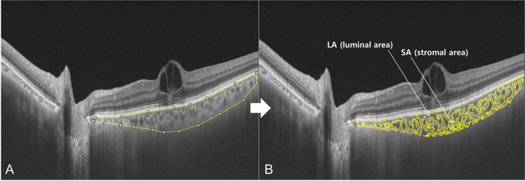
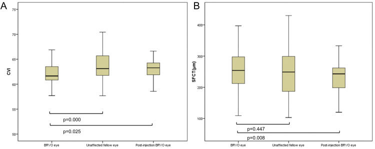
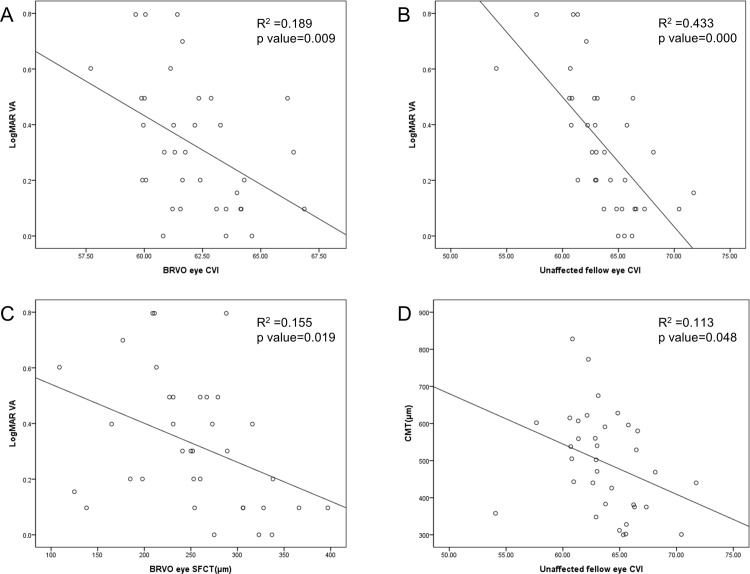
Methods: 35 patients with monocular BRVO and ME were analyzed retrospectively. Luminal and stromal areas in choroids of swept-source optical coherence tomography were calculated using the image binarization technique. The CVI was calculated as the ratio of the luminal to total choroidal area. The CVI of BRVO and ME eyes were compared with that of the unaffected fellow and post anti-vascular endothelial growth factor (VEGF) injected eyes. A regression analysis was performed on the choroidal parameters, logMAR visual acuity (VA) two years post disease onset and central macula thickness (CMT).
Results: The CVI of BRVO and ME eyes was significantly lower than the fellow and post-injected eyes (p<0.05). The regression analysis showed a strong association between two years after logMAR VA and the CVI of fellow eyes (R2 = 0.433, p<0.001). Remarkable correlations were observed in the CVI and subfoveal choroidal thickness of BRVO and ME eyes (R2 = 0.189, 0.155, respectively, p<0.05). The CMT of diseased eyes were also significantly associated with the CVI of unaffected fellow eyes (R2 = 0.113, p<0.05).
Conclusions: The alteration of CVI in BRVO and ME suggests that choroidal vasculature might be affected by extracellular fluid shift and VEGF changes. The fellow eye CVI could be a useful supplementary prognostic biomarker.
Conflict of interest statement
The authors have declared that no competing interests exist.
Figures
Similar articles
-
Longitudinal Assessment of the Choroidal Vascularity Index in Eyes with Branch Retinal Vein Occlusion-Associated Cystoid Macular Edema.Ophthalmol Ther. 2023 Aug;12(4):2103-2115. doi: 10.1007/s40123-023-00731-y. Epub 2023 May 23. Ophthalmol Ther. 2023. PMID: 37221425 Free PMC article.
-
The effect of branch retinal vein occlusion on the vascular structure of the choroid.Photodiagnosis Photodyn Ther. 2022 Mar;37:102687. doi: 10.1016/j.pdpdt.2021.102687. Epub 2021 Dec 17. Photodiagnosis Photodyn Ther. 2022. PMID: 34923154
-
Regional Choroidal Thickness Changes in Branch Retinal Vein Occlusion with Macular Edema.Ophthalmologica. 2015;234(2):109-18. doi: 10.1159/000437276. Epub 2015 Aug 19. Ophthalmologica. 2015. PMID: 26305536
-
Choroidal Perfusion Changes After Vitrectomy for Myopic Traction Maculopathy.Semin Ophthalmol. 2024 May;39(4):261-270. doi: 10.1080/08820538.2023.2283029. Epub 2023 Nov 21. Semin Ophthalmol. 2024. PMID: 37990380 Review.
-
Choroidal vascularity index: a step towards software as a medical device.Br J Ophthalmol. 2022 Feb;106(2):149-155. doi: 10.1136/bjophthalmol-2021-318782. Epub 2021 Jan 29. Br J Ophthalmol. 2022. PMID: 33514528 Review.
Cited by
-
Three-Dimensional Analysis of Choroidal Vessels in the Eyes of Patients With Unilateral BRVO.Front Med (Lausanne). 2022 Apr 5;9:854184. doi: 10.3389/fmed.2022.854184. eCollection 2022. Front Med (Lausanne). 2022. PMID: 35479961 Free PMC article.
-
Central and Peripheral Changes in Retinal Vein Occlusion and Fellow Eyes in Ultra-Widefield Optical Coherence Tomography Angiography.Invest Ophthalmol Vis Sci. 2024 Feb 1;65(2):6. doi: 10.1167/iovs.65.2.6. Invest Ophthalmol Vis Sci. 2024. PMID: 38306106 Free PMC article.
-
Radiomics Analysis Based on Optical Coherence Tomography to Prognose the Efficacy of Anti-VEGF Therapy of Retinal Vein Occlusion-Related Macular Edema.Invest Ophthalmol Vis Sci. 2025 Apr 1;66(4):74. doi: 10.1167/iovs.66.4.74. Invest Ophthalmol Vis Sci. 2025. PMID: 40277425 Free PMC article.
-
The role of fibronectin in aqueous humor and its association with retinal and choroidal vascular parameters in branch retinal vein occlusion.Graefes Arch Clin Exp Ophthalmol. 2025 May 24. doi: 10.1007/s00417-025-06819-4. Online ahead of print. Graefes Arch Clin Exp Ophthalmol. 2025. PMID: 40411584
-
Serial choriocapillaris flow changes in eyes with branched retinal vascular obstruction (BRVO).PLoS One. 2022 Nov 18;17(11):e0277988. doi: 10.1371/journal.pone.0277988. eCollection 2022. PLoS One. 2022. PMID: 36399455 Free PMC article.
References
-
- Noma H, Funatsu H, Yamasaki M, Tsukamoto H, Mimura T, Sone T, et al.. Pathogenesis of macular edema with branch retinal vein occlusion and intraocular levels of vascular endothelial growth factor and interleukin-6. Am J Ophthalmol. 2005. Aug;140(2):256–61. doi: 10.1016/j.ajo.2005.03.003 - DOI - PubMed
Publication types
MeSH terms
Substances
LinkOut - more resources
Full Text Sources
