. 2021 Oct 21;12(1):548.
doi: 10.1186/s13287-021-02614-0.

Hair follicle-derived mesenchymal stem cells decrease alopecia areata mouse hair loss and reduce inflammation around the hair follicle

Affiliations

Hair follicle-derived mesenchymal stem cells decrease alopecia areata mouse hair loss and reduce inflammation around the hair follicle

Weiyue Deng et al. Stem Cell Res Ther. .

Abstract

Background: Alopecia areata (AA) is a common autoimmune hair loss disease with increasing incidence. Corticosteroids are the most widely used for hair loss treatment; however, long-term usage of hormonal drugs is associated with various side effects. Mesenchymal stem cells (MSCs) therapy has been studied extensively to curb autoimmune diseases without affecting immunity against diseases.

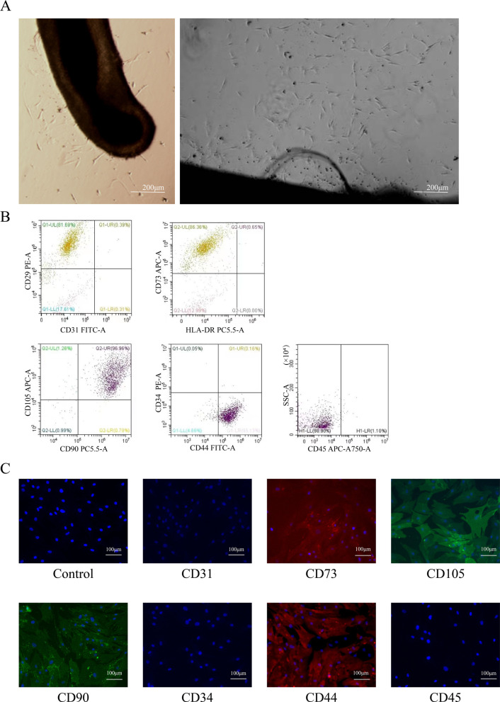
Methods: Hair follicle-derived MSCs (HF-MSCs) were harvested from the waste material of hair transplants, isolated and expanded. The therapeutic effect of HF-MSCs for AA treatment was investigated in vitro AA-like hair follicle organ model and in vivo C3H/HeJ AA mice model.

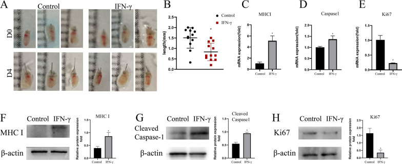
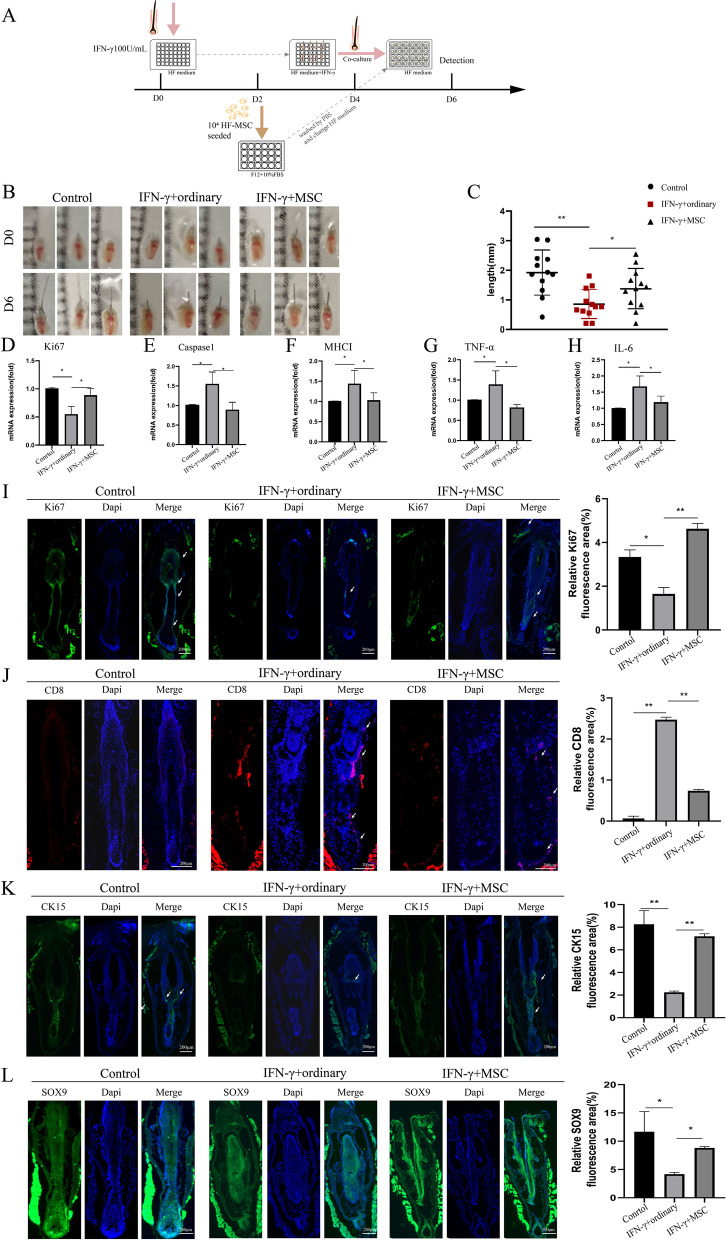
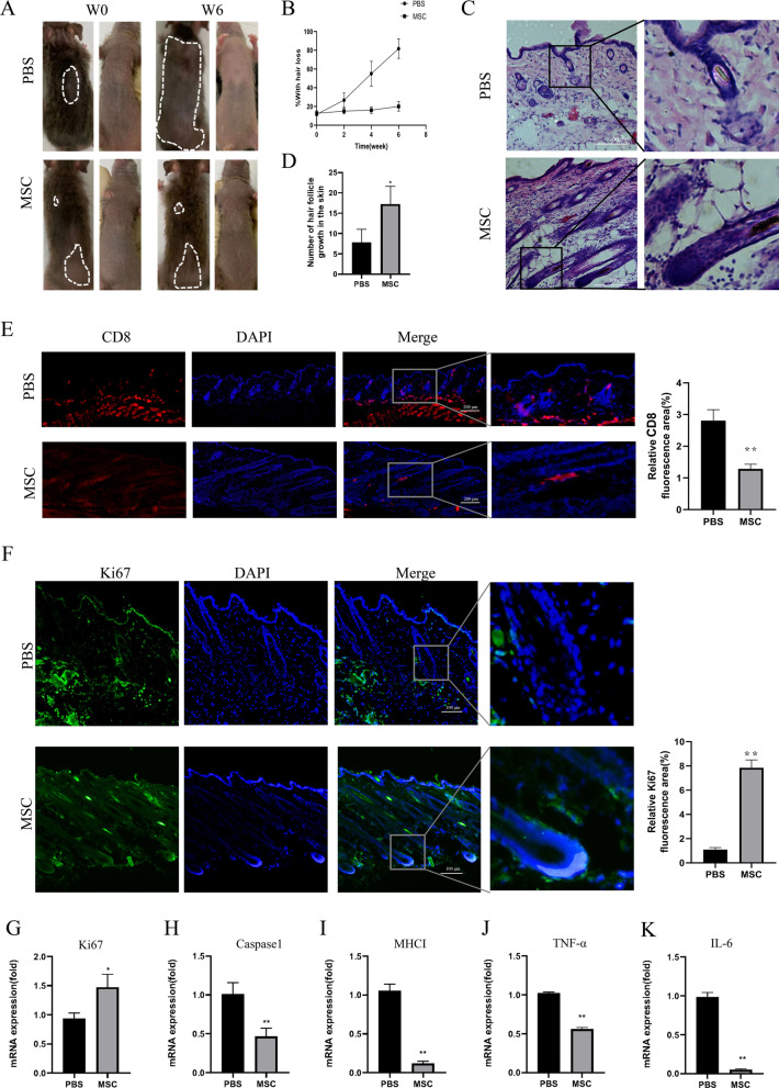
Results: AA-like hair follicle organ in vitro model was successfully established by pre-treatment of mouse vibrissa follicles by interferon-γ (IFN-γ). The AA-like symptoms were relieved when IFN-γ induced AA in vitro model was co-cultured with HF-MSC for 2 days. In addition, when skin grafted C3H/HeJ AA mice models were injected with 106 HF-MSCs once a week for 3 weeks, the transcription profiling and immunofluorescence analysis depicted that HF-MSCs treatment significantly decreased mouse hair loss and reduced inflammation around HF both in vitro and in vivo.

Conclusions: This study provides a new therapeutic approach for alopecia areata based on HF-MSCs toward its future clinical application.

Keywords: Alopecia areata; Hair follicle-derived mesenchymal stem cells; Hair loss treatment; Stem cell therapy.


Conflict of interest statement

The authors have no conflicting interests.

Figures

Fig. 1
Fig. 1
Isolation and characterization of human hair follicle-derived mesenchymal stem cells (HF-MSCs). A Cells that migrated from hair follicles and proliferated onto the culture plate exhibited fibroblast-like characteristics (bar = 200 μm). B Flow cytometry was used to measure the cell surface expression of MSC biomarkers. The cells expressed CD29, CD44, CD73, CD90, CD105 but not CD31, CD34, CD45, HLA-DR. C Immunofluorescence analysis was used to measure the surface markers of HF-MSCs. The cells expressed CD44, CD73, CD90, CD105 but not CD31, CD34, CD45 (bar = 100 μm)
Fig. 2
Fig. 2
Growth-suppression and MHC I expression upregulation of interferon (IFN)-γ treatment in the organ-cultured model. Mouse vibrissa follicles were isolated and cultured in HF medium with PBS or 100 IU mL−1 IFN-γ addition. A Mouse vibrissa follicles were photographed on day 0 and day 4. B Hair shaft growth was inhibited by interferon (IFN)-γ in organ-cultured mouse vibrissa follicles on the 4 days (n = 12/each group). C–E The mRNA transcript levels of MHC I, caspase1 and Ki67 in mouse vibrissa after 4 days of PBS or IFN-γ treatment measured by qRT-PCR, expression levels of those mRNA were normalized with GAPDH. F–H The protein expression levels of MHC I, caspase1 and Ki67 in mouse vibrissa after 4 days of PBS or IFN-γ treatment measured by western blotting. Relative protein expression levels were averaged from three groups of biology repeatedly and normalized with β-actin. The results were expressed as the Mean ± SD, *p < 0.05
Fig. 3
Fig. 3
Co-culture with HF-MSC suppressed inflammation, increased hair shaft growth in IFN-γ treated mouse vibrissa follicles. Two groups (n = 12/group) of Mouse vibrissa follicles had been isolated and cultured with 100 IU mL−1 IFN-γ for 4 days than both groups changed HF medium and one group co-culture with HF-MSC for 2 days. And one group was cultured in an ordinary HF medium over 6 days as a control. A Illustration of co-culture with HF-MSC in IFN-γ induced mouse vibrissa follicles in vitro model. Hair shafts were measured on day 0 and day 6. B–C Co-culture with HF-MSC significantly enhance hair shaft length (n = 12). D–H The mRNA transcript levels of Ki67, caspase1, MHC I, TNF-α, IL-6 in mouse vibrissa were examined using qRT-PCR on day 6, expression levels of those mRNA were normalized with GAPDH. I–L Immunofluorescence staining on day 6 of mouse vibrissa follicles (bar = 200 μm), co-culture with MSC increased Ki67 expression (green) (I), and suppressed CD8 expression (red) (J). IFN-γ intervention reduced CK15 (K) and SOX9 (L) markers expression levels, HF-MSC treatment reversed the reduction of those two markers. Relative fluorescence areas were averaged from 6 to 8 fields. The results were expressed as the Mean ± SD, *p < 0.05
Fig. 4
Fig. 4
Injection of HF-MSC suppressed AA in C3H/HeJ mice. A Hair loss in C3H/HeJ mice intravenously injection of HF-MSC weekly. At 0 and 6 weeks after injection, the image of mice were photographed. B Compared with injected PBS (n = 4), mice did not continue to lose his hair that injected HF-MSC (n = 5). C H&E showed that HF-MSC-injected mice exhibited more hair follicles in anagen, while the PBS group had dystrophic hairs with lymphocyte infiltration (bar = 100 μm).The skin samples were harvested after 6 weeks treatment. D The number of hair follicle growth in the skin, which was averaged from 6 slides (10×). EF HF-MSC-injection increased Ki67 expression (green) (bar = 100 μm) (E) and suppressed CD8 expression (red) (bar = 200 μm) (F) in C3H/HeJ skin. Relative fluorescence areas were averaged from 6 to 8 fields. G-K The mRNA transcript levels of caspase1, Ki67, MHC I, TNF-α, IL-6 in C3H/HeJ skin at 6 weeks after injection were examined using qRT-PCR, expression levels of those mRNA were normalized with GAPDH. The results were expressed as the Mean ± SD, *p < 0.05

References

    1. Al-Refu K. Stem cells and alopecia: a review of pathogenesis. Br J Dermatol. 2012;167:479–484. doi: 10.1111/j.1365-2133.2012.11018.x. - DOI - PubMed
    1. Lee S, Lee WS. Management of alopecia areata: updates and algorithmic approach. J Dermatol. 2017;44:1199–1211. doi: 10.1111/1346-8138.13933. - DOI - PubMed
    1. Safavi K. Prevalence of alopecia areata in the First National Health and Nutrition Examination Survey. Arch Dermatol. 1992;128:702. doi: 10.1001/archderm.1992.01680150136027. - DOI - PubMed
    1. Lee HH, Gwillim E, Patel KR, Hua T, Rastogi S, Ibler E, et al. Epidemiology of alopecia areata, ophiasis, totalis, and universalis: a systematic review and meta-analysis. J Am Acad Dermatol. 2020;82:675–682. doi: 10.1016/j.jaad.2019.08.032. - DOI - PubMed
    1. Toussi A, Barton VR, Le ST, Agbai ON, Kiuru M. Psychosocial and psychiatric comorbidities and health-related quality of life in alopecia areata: a systematic review. J Am Acad Dermatol. 2021;85:162–175. doi: 10.1016/j.jaad.2020.06.047. - DOI - PMC - PubMed

Publication types