. 2021 Oct 21;52(1):135.
doi: 10.1186/s13567-021-01005-w.

Glaesserella parasuis serotype 4 HPS4-YC disrupts the integrity of the swine tracheal epithelial barrier and facilitates bacterial translocation

Affiliations

Glaesserella parasuis serotype 4 HPS4-YC disrupts the integrity of the swine tracheal epithelial barrier and facilitates bacterial translocation

Qing Wang et al. Vet Res. .

Abstract

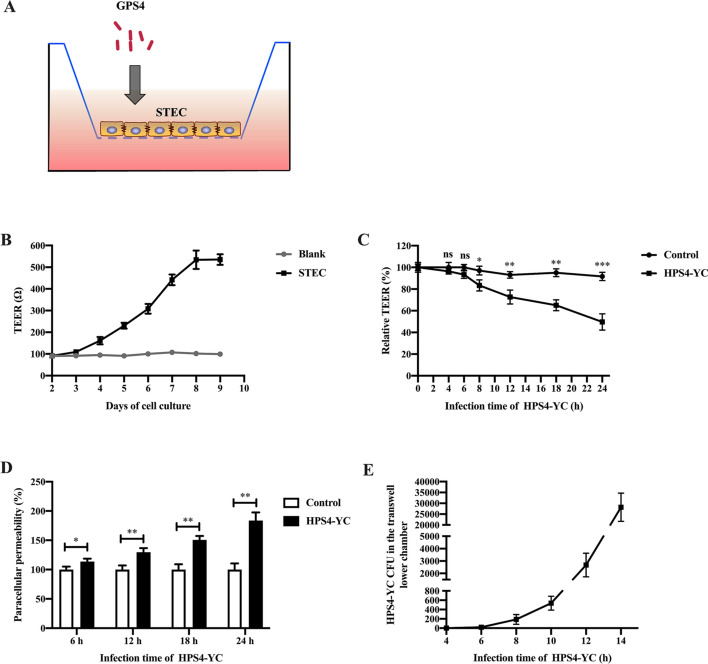
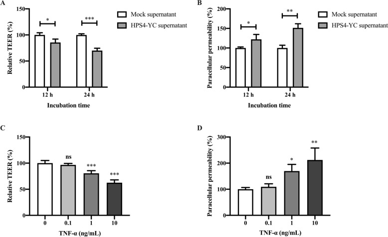
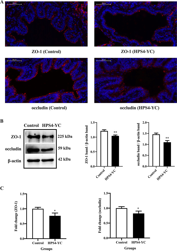
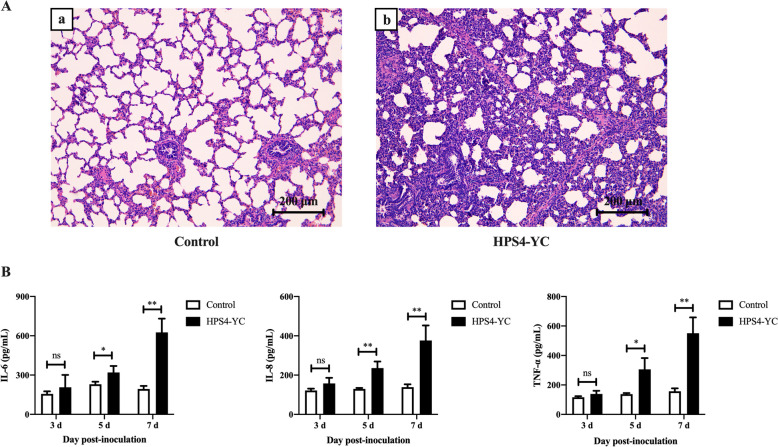
Glaesserella parasuis (G. parasuis) is a commensal bacterium in the upper respiratory tract of pigs that can also cause the swine Glässer disease, which induces an intensive inflammatory response and results in significant economic losses to the swine industry worldwide. G. parasuis can cause disease through infection of the respiratory tract, resulting in systemic infection, but the mechanism is largely unknown. Recently we showed that Glaesserella parasuis serotype 4 (GPS4) increased swine tracheal epithelial barrier permeability, resulting in easier bacterial translocation. Tight junction proteins (TJ) play a crucial role in maintaining the integrity and impermeability of the epithelial barrier. GPS4 decreased the expression of the TJ ZO-1 and occludin in swine tracheal epithelial cells (STEC). Furthermore, the proinflammatory cytokines IL-6, IL-8 and TNF-α were significantly upregulated in GPS4-infected STEC, and both the MAPK and NF-κB signaling pathways were activated and contributed to the expression of TNF-α. We demonstrate that the production of proinflammatory cytokines, especially TNF-α, during GPS4 infection was involved in barrier dysfunction. Additionally, animal challenge experiments confirmed that GPS4 infection downregulated TJ in the lungs of piglets and induced a severe inflammatory response. In general, G. parasuis infection downregulated the expression of TJ and induced massive secretion of proinflammatory cytokines, resulting in epithelial barrier disruption and favoring bacterial infection. This study allowed us to better understand the mechanism by which G. parasuis crosses the respiratory tract of pigs.

Keywords: Glaesserella parasuis; proinflammatory cytokines; swine tracheal epithelial cells; tight junction proteins.


Conflict of interest statement

The authors declare that they have no competing interests.

Figures

Figure 1
Figure 1
HPS4-YC infection in STEC. A Giemsa staining of HPS4-YC infecting STEC (100×). STEC were uninfected or infected with HPS4-YC at an MOI of 10, 100 or 1000 for 2 h. The black arrows point to the bacteria present. Scale bar, 20 μm. B, C Adherence and invasion assays. STEC were infected with HPS4-YC at different MOI for 2 h. The number of adherent and invasive bacteria per well was counted. D Cell viability of STEC were assessed by CCK-8. The results represent the means ± SD of three independent experiments. Significant differences in B and C were analyzed using Student t test. Significant differences in D were analyzed using one-way ANOVA. ns: not significant; ***P < 0.001.
Figure 2
Figure 2
HPS4-YC infection increased tracheal epithelial barrier permeability. A In vitro model of tracheal epithelial barrier. STEC were cultured in a transwell, GPS4 was added to the upper chamber. B Construction of an in vitro tracheal epithelial barrier model. C HPS4-YC infection decreased the TEER of the epithelial barrier. STEC monolayers were uninfected or infected with HPS4-YC for different times, and TEER was measured. D Measurement of paracellular permeability. The epithelial barrier integrity was analyzed by monitoring FITC-conjugated dextran crossing the transwell chamber. Paracellular permeability is shown relative to the uninfected permeability. E CFU of HPS4-YC across the epithelial barrier model were counted. The results are shown as the mean ± SD of three separate experiments. Significant differences in B and C were determined using the Student t test. ns: not significant; *, P < 0.05; **, P < 0.01; ***P < 0.001.
Figure 3
Figure 3
HPS4-YC infection induced disruption of TJ in STEC. STEC were infected with HPS4-YC for 6, 12, 18, and 24 h. A Protein levels of ZO-1 and occludin were analyzed by Western blot. The protein band intensities were quantified by ImageJ software. HPS4-YC caused a time-dependent decrease in tight junction protein levels in STEC. CT, the control group. B mRNA levels of ZO-1 and occludin were analyzed with qRT-PCR. The results are shown as the mean ± SD of triplicate independent experiments. Significant differences were analyzed using the Student t test. *, P < 0.05; **, P < 0.01; ***P < 0.001. C Immunofluorescence staining of ZO-1 and occludin in STEC. The infection times of HPS4-YC were 12 h and 24 h, then cells were fixed and the nucleus and TJ were stained (blue and green, respectively). HPS4-YC destroyed the integrity of TJ. Scale bar, 100 μm.
Figure 4
Figure 4
Proinflammatory cytokines were upregulated through activation of MAPK and NF-κB signaling pathways in HPS4-YC-infected STEC. A mRNA levels of cytokines IL-6, IL-8 and TNF-α were assessed through qRT-PCR assays. B Levels of the cytokines IL-6, IL-8 and TNF-α in culture supernatant of STEC were assessed by ELISA. C, D Activation of MAPK and NF-κB signaling pathways in STEC at 12 h and 24 h after HPS4-YC infection. The protein levels were determined by Western blot. E Expression of TNF-α was regulated by both MAPK and NF-κB signaling pathways. STEC were pretreated with DMSO or inhibitors for 1 h before HPS4-YC infection. The infection time of HPS4-YC was 12 h. SB203580, U0126, SP600125 and BAY11-7082 are inhibitors of p38, ERK, JNK and NF-κB, respectively. Data are shown as the mean ± SD of three independent experiments. Significant differences were determined using one-way ANOVA. *, P < 0.05; **, P < 0.01; ***P < 0.001.
Figure 5
Figure 5
Effect of inflammatory cytokines on swine tracheal epithelial barrier damage. A, B Cytokines present in the supernatant decreased TEER and increased paracellular permeability respectively, promoting barrier damage. The supernatant of HPS4-YC-infected and mock-infected STEC was added to the upper chambers of transwells, and TEER and FITC-conjugated dextran crossing the transwell chamber were measured at 12 h and 24 h. C, D TNF-α damaged the epithelial barrier integrity. Different concentrations of TNF-α were added to the medium in the apical compartment for 24 h, and TEER and paracellular permeability were measured to determine their effect on epithelial barrier integrity. Data are shown as the mean ± SD of triplicate assays. Significant differences in A and B were determined using Student t test. Significant differences in C and D were determined using one-way ANOVA. ns: not significant; *, P < 0.05; **, P < 0.01; ***P < 0.001.
Figure 6
Figure 6
HPS4-YC downregulated tight junction protein levels in the lungs of piglets. A Immunofluorescence staining of ZO-1 and occludin in the lungs. Blue, the nucleus. Red, ZO-1 or occludin. Scale bar, 50 μm. B Protein levels of ZO-1 and occludin in the lungs were analyzed by Western blot. HPS4-YC decreased the expression of ZO-1 and occludin. C mRNA levels of ZO-1 and occludin in the lungs were analyzed with qRT-PCR. The results were in accordance with cell experiments. Data are shown as the mean ± SD of three independent experiments. Significant differences were determined using the Student t test. *, P < 0.05; **, P < 0.01.
Figure 7
Figure 7
Histopathological examination of the lungs and detection of proinflammatory cytokines in serum. A Representative histopathological images of piglets mock-infected or infected with HPS4-YC. Scale bar, 200 μm. B Levels of the proinflammatory cytokines IL-6, IL-8 and TNF-α in serum samples were assessed by ELISA. Data are presented as mean ± SD of three independent experiments. Significant differences were determined using the Student t test. ns not significant; *, P < 0.05; **, P < 0.01.

References

    1. Hau SJ, Eberle KC, Brockmeier SL. Importance of strain selection in the generation of heterologous immunity to Glaesserella (Haemophilus) parasuis. Vet Immunol Immunopathol. 2021;234:110205. doi: 10.1016/j.vetimm.2021.110205. - DOI - PubMed
    1. Costa-Hurtado M, García-Rodríguez L, López-Serrano S, Aragón V. Haemophilus parasuis VtaA2 is involved in adhesion to extracellular proteins. Vet Res. 2019;50:69. doi: 10.1186/s13567-019-0687-2. - DOI - PMC - PubMed
    1. Jia A, Zhou R, Fan H, Yang K, Zhang J, Xu Y, Wang G, Liao M. Development of serotype-specific PCR assays for typing of Haemophilus parasuis isolates circulating in southern China. J Clin Microbiol. 2017;55:3249–3257. doi: 10.1128/JCM.00688-17. - DOI - PMC - PubMed
    1. Macedo N, Rovira A, Torremorell M. Haemophilus parasuis: infection, immunity and enrofloxacin. Vet Res. 2015;46:128. doi: 10.1186/s13567-015-0263-3. - DOI - PMC - PubMed
    1. Costa-Hurtado M, Barba-Vidal E, Maldonado J, Aragón V. Update on Glasser's disease: how to control the disease under restrictive use of antimicrobials. Vet Microbiol. 2020;242:108595. doi: 10.1016/j.vetmic.2020.108595. - DOI - PubMed

MeSH terms

LinkOut - more resources