. 2021 Nov 1;35(21-22):1431-1444.
doi: 10.1101/gad.348797.121. Epub 2021 Oct 21.

Neural stem/precursor cells dynamically change their epigenetic landscape to differentially respond to BMP signaling for fate switching during brain development

Affiliations

Neural stem/precursor cells dynamically change their epigenetic landscape to differentially respond to BMP signaling for fate switching during brain development

Sayako Katada et al. Genes Dev. .

Abstract

During neocortical development, tight regulation of neurogenesis-to-astrogenesis switching of neural precursor cells (NPCs) is critical to generate a balanced number of each neural cell type for proper brain functions. Accumulating evidence indicates that a complex array of epigenetic modifications and the availability of extracellular factors control the timing of neuronal and astrocytic differentiation. However, our understanding of NPC fate regulation is still far from complete. Bone morphogenetic proteins (BMPs) are renowned as cytokines that induce astrogenesis of gliogenic late-gestational NPCs. They also promote neurogenesis of mid-gestational NPCs, although the underlying mechanisms remain elusive. By performing multiple genome-wide analyses, we demonstrate that Smads, transcription factors that act downstream from BMP signaling, target dramatically different genomic regions in neurogenic and gliogenic NPCs. We found that histone H3K27 trimethylation and DNA methylation around Smad-binding sites change rapidly as gestation proceeds, strongly associated with the alteration of accessibility of Smads to their target binding sites. Furthermore, we identified two lineage-specific Smad-interacting partners-Sox11 for neurogenic and Sox8 for astrocytic differentiation-that further ensure Smad-regulated fate-specific gene induction. Our findings illuminate an exquisite regulation of NPC property change mediated by the interplay between cell-extrinsic cues and -intrinsic epigenetic programs during cortical development.

Keywords: epigenetic modifications; Smad; astrocyte; bone morphogenetic protein; chromatin accessibility; differentiation; neural stem/precursor cell; neuron.


Figures

Figure 1.
Figure 1.
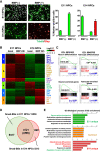
BMP2 induces neuronal and astrocytic differentiation in mid- and late-gestational NPCs, respectively. (A) NPCs derived from E11 or E14 forebrain were differentiated for 4 d in the presence or absence of BMP2, and immunostained with anti-Tubb3 (green) and anti-Gfap (red) antibodies. (B) RNA-seq was performed on E11- and E14-derived NPCs before and 24 h after BMP2 treatment. Heat maps for the top 2500 variable genes are shown together with the results of K-means clustering using k = 6. The colors display the relative gene expression on a log2 scale. Green indicates the lowest, black indicates the intermediate, and red indicates the highest expression. (C) Enrichment score plots for neuron differentiation (GO: 0030182), astrocyte differentiation (GO: 0048708), and neuron- or astrocyte-enriched gene sets using BMP2-treated E11 versus E14 NPCs. (D) Venn diagram showing BMP2-induced Smad-BSs in E11 and E14 NPCs identified by ChIP-seq. (E) GO terms (biological process) for E11 unique, E14 unique, and E11/14 common Smad-BS-associated genes.
Figure 2.
Figure 2.
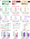
Chromatin accessibility regulates developmental stage-specific Smad binding. (A) Bar graphs showing the number of Smad-BSs located within 20 kb from the nearest TSS. Associated genes were further classified based on expression changes using RNA-seq data. Besides up-regulated (Up) and down-regulated (Down) genes, common peaks have an “either” category, since some genes exhibit opposite induction in E11 and E14 NPCs following BMP2 stimulation. (B) Plots of Smad ChIP-seq signals around E11 unique, E14 unique, and E11/14 common Smad-BSs. Three types of Smad-BSs were further classified based on their expression change, up-regulated or down-regulated, and either for E11/E14 common Smad-BSs. Smad enrichment profiles of unchanged genes were also plotted (Unch.; dashed lines). (C) Plots of ATAC-seq signals around E11 unique, E14 unique, and E11/14 common Smad-BSs in E11-derived (blue) or E14-derived (magenta) NPCs. (D) Representative genes having E11 unique, E14 unique, and E11/14 common Smad-BSs. Tracks display ATAC-seq signals of basal E11 (blue) and E14 (magenta) NPCs, or Smad ChIP-seq of basal or BMP2-treated E11 (green) and E14 (red) NPCs. Signal enrichment for the boxed regions is expanded in the bar graphs.
Figure 3.
Figure 3.
Epigenetic modifications regulate stage-specific Smad binding. (A) Plots of H3K27me3 ChIP-seq signals around E11 unique, E14 unique, and E11/14 common Smad-BSs in E11-derived (blue) or E14-derived (orange) NPCs. (B) Averaged DNA methylation levels of all CpG sites located within E11 unique (n = 60,813), E14 unique (n = 1353), and E11/14 common (n = 48,924) Smad-BSs in NPCs directly isolated from E11, E14, and E18 mouse forebrains. (C) Representative genes having proximal coincident R-DMRs and E14 unique Smad-BSs. Tracks display bisulfite-seq of E11 and E18 NPCs, ATAC-seq of E11 (blue) and E14 (magenta) NPCs, and Smads ChIP-seq of untreated or BMP2-treated E14 NPCs (red). Black lines at the bottom show R-DMRs. (D) E11 NPCs were mock-infected or infected with Nfia-expressing virus and cultured for 2 d in the presence of bFGF, and then stimulated with BMP2 for a further 4 d to induce differentiation. The cells were stained with antibodies against Tubb3 (green) and Gfap (red). (E) E11 NPCs were mock-infected (black) or infected with Nfia-expressing virus (red) and cultured for 2 d in the presence of bFGF, and mRNA was isolated before and 24 h after BMP2 stimulation. Real-time RT-PCR data for selected genes are shown as means ± SD (n = 3).
Figure 4.
Figure 4.
Smads interact with SoxC and SoxE family proteins and co-occupy Smad-BSs. (A) Classification of Smad-BSs identified in this study or reported in mesodermal cells based on chromatin state in NPCs. Chromatin openness was categorized into four states—E11 uniquely open (blue), E14 uniquely open (red), E11/14 commonly open (magenta), or none of these (black)—using ATAC-seq data. (B,left) Venn diagrams showing the number and percentage of Smad co-occupancy in Sox4- or Sox9-bound sites. (Right) Sox4/9 and Smad cobound sites were divided into either E11 unique (green), E14 unique (red), or E11/14 common (orange) Smad-BSs as shown in the pie charts. (C) Myc-tagged Smad1 and Smad4 were transfected with HA-tagged Sox family proteins into HEK293T cells together with ALK3-QD and immunoprecipitated using anti-HA antibody. Nonspecific bands observed in the HA blot are indicated by an asterisk. (D) Lysates of BMP2-stimulated E11 or E14 NPCs were precipitated by anti-phospho-Smad1/5/9 antibody and blotted with the indicated antibodies.
Figure 5.
Figure 5.
Smad-interacting partners (Sox11 for neurogenic and Sox8 for astrocytic differentiation) ensure Smad-regulated fate-specific gene induction. (A) Expression levels of SoxB1/C/E family genes in E11 or E14 NPCs are depicted as a heat map. (B) E14-derived NPCs were infected with lentivirus engineered to express Sox11 and cultured for 2 d with bFGF, and mRNA was isolated before and 24 h after BMP2 stimulation. Real-time RT-PCR data are show as means ± SD (n = 3). (**) P < 0.01, (*) P < 0.05 by one-way ANOVA with Bonferroni's multiple comparison test. (C) E11-derived NPCs were infected with lentivirus to express Sox8 (red) or control (black) and cultured for 2 d with bFGF, and mRNA was isolated before and 24 h after BMP2 treatment. Real-time RT-PCR data are shown as means ± SD (n = 3). (D) Luciferase assay of Gfap promoter in E14-derived NPCs. Together with the reporter, Sox8 or Sox11 were cotransfected as indicated, and the cells were stimulated with BMP2 for 24 h (gray). Relative light units compared with control without BMP2 (white) are indicated. Bar graph shows means ± SD (n = 4). (**) P < 0.01, (*) P < 0.05 by one-way ANOVA with Bonferroni's multiple comparison test.
Figure 6.
Figure 6.
Schematic model for the effects of BMPs on developmentally different NPCs. In mid-gestation, chromatin accessibility of astrocytic genes is restricted by epigenetic modifications such as DNA methylation and H3K27 trimethylation; however, during NPC progression, these repressive epigenetic marks are removed and, in turn, neurogenic genes acquire H3K27 trimethylation, leading to a less accessible chromatin state. Upon BMP stimulation, activated Smads bind to reachable consensus sequences, but developmental stage-specific Smad partners further ensure Smad-regulated fate-specific gene induction.

Similar articles

Cited by

References

    1. Akizu N, Estarás C, Guerrero L, Martí E, Martínez-Balbás MA. 2010. H3k27me3 regulates BMP activity in developing spinal cord. Development 137: 2915–2925. 10.1242/dev.049395 - DOI - PubMed
    1. Albert M, Huttner WB. 2018. Epigenetic and transcriptional pre-patterning—an emerging theme in cortical neurogenesis. Front Neurosci 12: 359. 10.3389/fnins.2018.00359 - DOI - PMC - PubMed
    1. Baik J, Magli A, Tahara N, Swanson SA, Koyano-Nakagawa N, Borges L, Stewart R, Garry DJ, Kawakami Y, Thomson JA, et al. 2016. Endoglin integrates BMP and Wnt signalling to induce haematopoiesis through JDP2. Nat Commun 7: 13101. 10.1038/ncomms13101 - DOI - PMC - PubMed
    1. Barnabé-Heider F, Wasylnka JA, Fernandes KJL, Porsche C, Sendtner M, Kaplan DR, Miller FD. 2005. Evidence that embryonic neurons regulate the onset of cortical gliogenesis via cardiotrophin-1. Neuron 48: 253–265. 10.1016/j.neuron.2005.08.037 - DOI - PubMed
    1. Bonaguidi MA, McGuire T, Hu M, Kan L, Samanta J, Kessler JA. 2005. LIF and BMP signaling generate separate and discrete types of GFAP-expressing cells. Development 132: 5503–5514. 10.1242/dev.02166 - DOI - PubMed

Publication types

Substances