. 2021 Oct 26;118(43):e2113852118.
doi: 10.1073/pnas.2113852118.

Serotonin modulates melatonin synthesis as an autocrine neurotransmitter in the pineal gland

Affiliations

Serotonin modulates melatonin synthesis as an autocrine neurotransmitter in the pineal gland

Bo Hyun Lee et al. Proc Natl Acad Sci U S A. .

Abstract

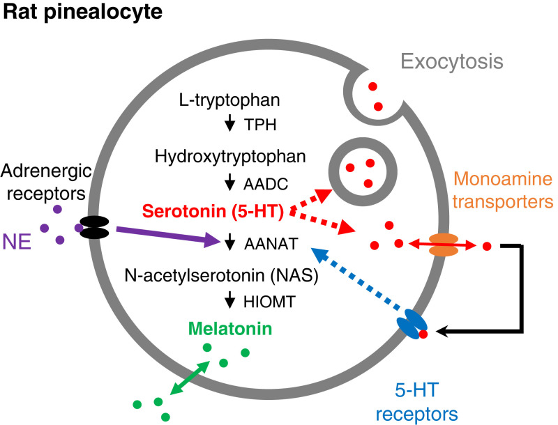
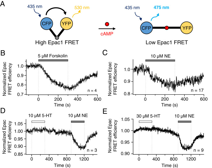
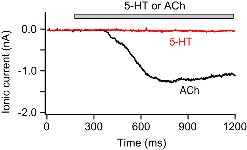
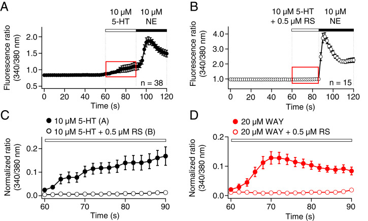
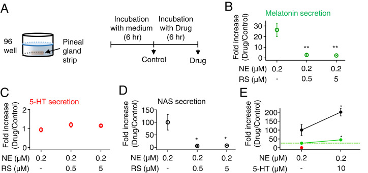
The pineal gland secretes melatonin principally at night. Regulated by norepinephrine released from sympathetic nerve terminals, adrenergic receptors on pinealocytes activate aralkylamine N-acetyltransferase that converts 5-hydroxytryptamine (5-HT, serotonin) to N-acetylserotonin, the precursor of melatonin. Previous studies from our group and others reveal significant constitutive secretion of 5-HT from pinealocytes. Here, using mass spectrometry, we demonstrated that the 5-HT is secreted primarily via a decynium-22-sensitive equilibrative plasma membrane monoamine transporter instead of by typical exocytotic quantal secretion. Activation of the endogenous 5-HT receptors on pinealocytes evoked an intracellular Ca2+ rise that was blocked by RS-102221, an antagonist of 5-HT2C receptors. Applied 5-HT did not evoke melatonin secretion by itself, but it did potentiate melatonin secretion evoked by submaximal norepinephrine. In addition, RS-102221 reduced the norepinephrine-induced melatonin secretion in strips of pineal gland, even when no exogenous 5-HT was added, suggesting that the 5-HT that is constitutively released from pinealocytes accumulates enough in the tissue to act as an autocrine feedback signal sensitizing melatonin release.

Keywords: N-acetylserotonin; melatonin; pineal gland; plasma membrane monoamine transporter; serotonin receptor.


Conflict of interest statement

The authors declare no competing interest.

Figures

Fig. 1.
Fig. 1.
Synthesis and release of 5-HT and melatonin in rat pinealocytes. The enzymatic pathway from L-tryptophan to 5-HT and melatonin uses four enzymes. AANAT is regulated by adrenergic signaling. Two possibilities are shown (red dotted arrows) for the mechanism of release of 5-HT from pinealocytes: through vesicular exocytosis or through monoamine transporters running in reverse (orange). Released serotonin activates serotonin receptors (blue) on the plasma membrane of pinealocytes. We are suggesting that the receptors up-regulate AANAT acting as an auto- or paracrine mechanism to promote melatonin synthesis. AADC, aromatic amino acid decarboxylase; HIOMT, hydroxyindole-O-methyltransferase; TPH, tryptophan hydroxylase.
Fig. 2.
Fig. 2.
NE promotes an early transient 5-HT release. (A) A single PG from a rat was sliced into three to four slices (400 μm thick), and each slice was further sliced into three to five strips (300 to 400 μm thick). (B) Schematic drawing of a single pineal strip in one well of a 96-well plate. Individual strips were incubated in M199 for 30 min and then in M199 (Control) or M199 plus 1 μM NE or 50 μM ACh for another 40 min. After each incubation, the medium was collected and used to measure 5-HT (C), NAS (D), and melatonin (E) levels by UPLC/MS analysis. Fold increases are given as test period divided by baseline period and compared using a t test. **P < 0.01. Results are presented as mean ± SEM.
Fig. 3.
Fig. 3.
Serotonin is not released via exocytosis. Representative amperometric current traces measured using carbon-fiber amperometry with a miniature amperometric electrode polarized at +600 mV. (A) Amperometric traces of pinealocytes preloaded with exogenous dopamine. Enlarged single quantal events in the Right are from the numbered spikes from the Left (1: spontaneous event, 2, 3: evoked exocytosis). The three amperometric spikes illustrated correspond to 2-electron oxidation of 1.7 × 10⁷, 2.1 × 10⁷, and 270 × 107 dopamine molecules, respectively. In the remaining panels, pinealocytes were not preloaded with dopamine. The amperometric electrode was placed in the bath solution without pinealocytes (control traces) (B and D) or on a cluster of pinealocytes (C and E). ACh (50 μM) and high potassium chloride (70 mM KCl) were used to elicit membrane depolarization and intracellular Ca2+ increases. Next, 1 μM NE (an oxidizable molecule) was applied to the electrode in the bath (D) or with the same electrode touching pinealocytes (E). No quantal events were observed before or after NE application, only a step up of current as applied NE is being oxidized.
Fig. 4.
Fig. 4.
5-HT is released from pineal strips via a decynium-sensitive plasma membrane monoamine transporter. (A) Schematic of a single strip inserted into a microchamber connected to a peristaltic pump for continuous perifusion. (B) Time course of 5-HT secretion during drug exposure from UPLC/MS analysis of 20-min samples of the solution passing through the pineal strip. Open circles are control and closed circles are the secretion in the presence of 50 μM decynium-22, a selective inhibitor of equilibrative cation transporters, applied only during drug periods D1, D2, and D3.
Fig. 5.
Fig. 5.
5-HT does not change cAMP levels in isolated pinealocytes. The cAMP levels were analyzed by FRET. (A) A schematic diagram of the cAMP biosensor, Epac1, sandwiched between CFP (donor) and YFP (acceptor). Pinealocytes were transfected with the Epac1 probe by magnetofection. Once cAMP binds to Epac1, the conformational change increases the distance between CFP and YFP and thereby decreases FRET efficiency. (B) Forskolin, increasing cAMP, lowers FRET efficiency in pinealocytes. (C) NE activates endogenous β-adrenergic receptors and increases cAMP levels in pinealocytes. Neither 10 μM 5-HT (D) nor 30 μM 5-HT (E) affected the level of cAMP in pinealocytes.
Fig. 6.
Fig. 6.
5-HT does not activate an ionotropic 5-HT3 receptor in pinealocytes. Representative recordings of 5-HT– and ACh-induced currents from a pinealocyte in whole-cell patch-clamp recording. Six of six cells showed no current with 5-HT and all of them exhibited significant inward currents with ACh. Holding potential was −60 mV. ACh (50 μM) and 5-HT (30 μM) were applied to the cell during the bar using local perfusion.
Fig. 7.
Fig. 7.
5-HT activates 5-HT2C receptors in the membrane of pinealocytes and raises calcium. Intracellular calcium levels were monitored by calcium imaging using Fura-2 AM fluorescence ratios (340/380 nm). (A) Calcium time course during application of 5-HT and NE. (B) Calcium time course in pinealocytes preincubated for 10 min with 0.5 μM RS, a selective 5-HT2C antagonist, and then measured during application of 5-HT with RS and NE. (C) Magnified traces from the red-dashed box (A and B), showing that RS blocked the induction of a modest calcium increase by 5-HT. Closed circles indicate the application of 5-HT (A) and open circles indicate coapplication of 5-HT and RS (B). (D) Like 5-HT, WAY, a potent and selective 5-HT2C agonist, increased intracellular calcium by a pathway blocked by RS.
Fig. 8.
Fig. 8.
5-HT2C receptors sensitize NE-induced melatonin and NAS secretion in pinealocytes. (A) Schematic of a single pineal strip in one well of a 96-well plate. Strips were incubated individually in M199 for 6 h and then in M199 containing different drugs for another 6 h. Next, 0.2 μM NE was used to activate adrenergic receptors, and RS was used to block 5-HT2C receptors. After each 6-h incubation, the solutions were collected and used to measure melatonin (B), 5-HT (C), and NAS (D) levels by UPLC/MS. (E) Normalized levels of secreted melatonin (green), 5-HT (red), and NAS (black) after further application of 5-HT with NE were measured as a positive control. The green dotted line indicates the basal melatonin secretion. When exogenous 5-HT was added to the bath, it was not possible to measure endogenous 5-HT secretion. Fold increases relative to the baseline incubation in M199 were compared using a t test. **P < 0.01 and *P < 0.05.

Similar articles

Cited by

References

    1. Klein D. C., et al. ., The melatonin rhythm-generating enzyme: Molecular regulation of serotonin N-acetyltransferase in the pineal gland. Recent Prog. Horm. Res. 52, 307–357, discussion 357–358 (1997). - PubMed
    1. Sun X., Liu T., Deng J., Borjigin J., Long-term in vivo pineal microdialysis. J. Pineal Res. 35, 118–124 (2003). - PubMed
    1. Møller M., Baeres F. M., The anatomy and innervation of the mammalian pineal gland. Cell Tissue Res. 309, 139–150 (2002). - PubMed
    1. Masson J., Emerit M. B., Hamon M., Darmon M., Serotonergic signaling: Multiple effectors and pleiotropic effects. Wiley Interdiscip. Rev. Membr. Transp. Signal. 1, 685–713 (2012).
    1. Ray R. S., et al. ., Impaired respiratory and body temperature control upon acute serotonergic neuron inhibition. Science 333, 637–642 (2011). - PMC - PubMed

Publication types

LinkOut - more resources