

Interthalamic adhesion in humans: a gray commissure?
- PMID: 34675136
- PMCID: PMC8968232
- DOI: 10.5115/acb.21.164
Interthalamic adhesion in humans: a gray commissure?
Abstract
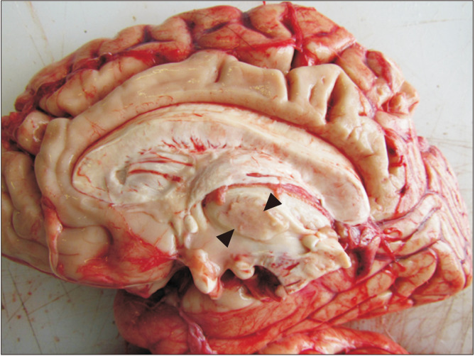
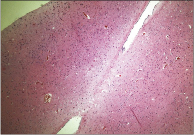
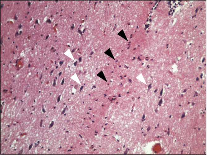
Interthalamic adhesion is an inconstant part of the human diencephalic neuroanatomy, which some histological studies have indicated it is a gray commissure and others a white commissure. Its presence has been associated with alterations in health status, including schizophrenia, psychotic states, and hydrocephalus. Thirty-one fresh human brains were evaluated randomly, to determine the presence of interthalamic adhesion and its histological composition, by way of lamina terminalis puncture of the third ventricle. Photographic records were taken and histological processes was performed by hematoxylin-eosin staining, in the case of the existence of the adhesion. It was found that 51.71% did present interthalamic adhesion, and on histological examination, no neuron bodies were found in the median part, which implies that does not correspond to a gray commissure, but interthalamic adhesion in humans is variable, with a predominance of glial cells. There is no gray commissure in human interthalamic adhesions.
Keywords: Anatomy; Diencephalon; Histology; Neuroanatomy; Thalamus.
Conflict of interest statement
No potential conflict of interest relevant to this article was reported.
Figures
References
-
- Federative Committee on Anatomical Terminology, author. Terminologia anatomica: international anatomical terminology. Thieme; New York: 1998. p. 120.
-
- Malobabić S, Puskas L, Blagotić M. Size and position of the human adhaesio interthalamica. Gegenbaurs Morphol Jahrb. 1987;133:175–80. - PubMed
-
- Trzesniak C, Kempton MJ, Busatto GF, de Oliveira IR, Galvão-de Almeida A, Kambeitz J, Ferrari MC, Filho AS, Chagas MH, Zuardi AW, Hallak JE, McGuire PK, Crippa JA. Adhesio interthalamica alterations in schizophrenia spectrum disorders: a systematic review and meta-analysis. Prog Neuropsychopharmacol Biol Psychiatry. 2011;35:877–86. doi: 10.1016/j.pnpbp.2010.12.024. - DOI - PubMed
LinkOut - more resources
Full Text Sources