. 2021 Oct 21;11(1):20854.
doi: 10.1038/s41598-021-00229-2.

DNA damage response of haematopoietic stem and progenitor cells to high-LET neutron irradiation

Affiliations

DNA damage response of haematopoietic stem and progenitor cells to high-LET neutron irradiation

Monique Engelbrecht et al. Sci Rep. .

Abstract

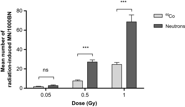
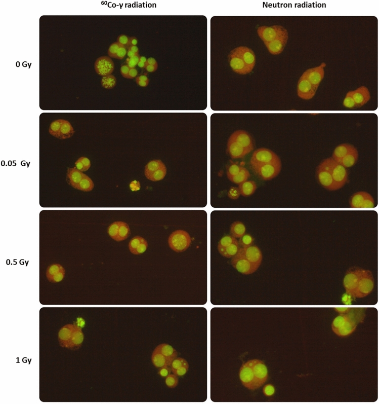
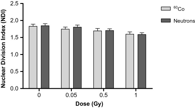
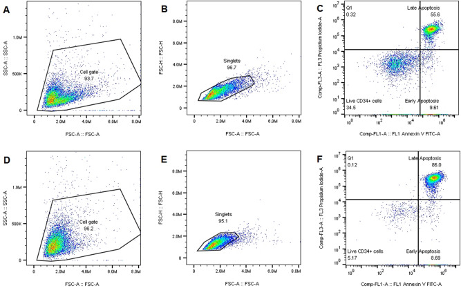
The radiosensitivity of haematopoietic stem and progenitor cells (HSPCs) to neutron radiation remains largely underexplored, notwithstanding their potential role as target cells for radiation-induced leukemogenesis. New insights are required for radiation protection purposes, particularly for aviation, space missions, nuclear accidents and even particle therapy. In this study, HSPCs (CD34+CD38+ cells) were isolated from umbilical cord blood and irradiated with 60Co γ-rays (photons) and high energy p(66)/Be(40) neutrons. At 2 h post-irradiation, a significantly higher number of 1.28 ± 0.12 γ-H2AX foci/cell was observed after 0.5 Gy neutrons compared to 0.84 ± 0.14 foci/cell for photons, but this decreased to similar levels for both radiation qualities after 18 h. However, a significant difference in late apoptosis was observed with Annexin-V+/PI+ assay between photon and neutron irradiation at 18 h, 43.17 ± 6.10% versus 55.55 ± 4.87%, respectively. A significant increase in MN frequency was observed after both 0.5 and 1 Gy neutron irradiation compared to photons illustrating higher levels of neutron-induced cytogenetic damage, while there was no difference in the nuclear division index between both radiation qualities. The results point towards a higher induction of DNA damage after neutron irradiation in HSPCs followed by error-prone DNA repair, which contributes to genomic instability and a higher risk of leukemogenesis.


Conflict of interest statement

The authors declare no competing interests.

Figures

Figure 1
Figure 1
Mean number of micronuclei (MN) in CD34+ cells (n = 12) induced by different doses (0.05, 0.5 and 1 Gy) of 60Co γ-rays and p(66)/Be(40) neuron irradiation. The number of MN induced by the irradiation was obtained by subtracting the mean number of MN in the non-irradiated controls (0 Gy) from the mean MN number scored in the irradiated samples. MN yields were significantly higher post-neutron irradiation compared to 60Co γ-rays (***p < 0.001) for the 0.5 Gy and 1 Gy dose (***p < 0.001) but not for the 0.05 Gy dose (ns). Error bars represent the standard error of the mean (SEM) of the 12 different donors for each radiation quality. At least 1000 BN cells were scored for each donor per condition. No significant difference is indicated by ns.
Figure 2
Figure 2
Characteristic appearance of human CD34+ cells for the different radiation doses that were investigated as part of the CBMN assay. The images illustrate the BN CD34+ cells containing micronuclei after low-LET 60Co γ-rays (left panel) and high-LET neutrons (right panel) exposure. The images were captured from a fluorescent Zeiss Axio Imager A1 microscope, at 20× magnification.
Figure 3
Figure 3
The nuclear division index (NDI) was calculated to compare the proliferation status of the micro-culture CBMN assay for the CD34+ samples irradiated with different radiation qualities with doses of 0, 0.05, 0.5 and 1 Gy. Error bars represent the standard error of the mean (SEM) of the 12 different donors for each radiation quality. At least 500 viable cells were manually scored for each donor per condition.
Figure 4
Figure 4
Mean number of radiation-induced γ-H2AX foci per CD34+ cell at 2 and 18 h post-irradiation with 0.5 Gy. The number of radiation-induced γ-H2AX foci was obtained by subtracting the mean number of γ-H2AX foci in the non-irradiated controls from the mean γ-H2AX foci number scored in the irradiated samples. The number of radiation-induced γ-H2AX foci was significantly different between 60Co γ-rays (n = 6) and neutron (n = 9) radiation at 2 h (*p < 0.05), while after 18 h no significant difference (p > 0.05) was observed between the two radiation qualities. Error bars represent standard error of the mean (SEM).The images (AD) on the right-side show immunofluorescence staining of γ-H2AX foci with a TRITC-conjugated secondary antibody (red). The foci represent the residual DNA DSBs at 2 h (A and C) and 18 h (B and D) after 0.5 Gy 60Co γ-rays (A and B) and neutron irradiation (C and D).
Figure 5
Figure 5
The gating strategy for the Annexin-V/PI apoptosis analysis. CD34+ cells were gated on forward (FSC) versus side scatter (SSC) to select the cell population (A and D). Next, the cells were gated on FSC-Height (FSC-H) vs FSC-Area (FSC-A) to exclude all the doublets and to generate the singlets gate (B and E). Finally, all the subpopulations were analysed on the Annexin V-FITC versus PI scatter for live, early and late apoptosis at 18 h post-irradiation (C and F). The upper part (AC) represents the gating strategy of CD34+ cells irradiated with a low dose of neutrons at 0.5 Gy; and the lower part (DF) CD34+ cells irradiated with a high dose at 3 Gy which resulted in a higher percentage of late apoptosis.
Figure 6
Figure 6
The spread in purity of the CD34+ samples as measured with the BD Accuri™ C6 flow cytometer. The average purity of the CD34+ cells was 94.11%. The error bar represents the standard error of the mean (SEM) of the different isolated samples (n = 34).

References

    1. Britel M, Bourguignon M, Foray N. The use of the term ‘radiosensitivity’ through history of radiation: From clarity to confusion. Int. J. Radiat. Biol. 2018;94:503–512. doi: 10.1080/09553002.2018.1450535. - DOI - PubMed
    1. Shao L, Luo Y, Zhou D. Hematopoietic stem cell injury induced by ionizing radiation. Antioxid. Redox Signal. 2014;20:1447–1462. doi: 10.1089/ars.2013.5635. - DOI - PMC - PubMed
    1. Doulatov S, Notta F, Laurenti E, Dick JE. Hematopoiesis: A human perspective. Cell Stem Cell. 2012;10:120–136. doi: 10.1016/j.stem.2012.01.006. - DOI - PubMed
    1. Pennings S, Liu KJ, Qian H. The stem cell niche: Interactions between stem cells and their environment. Stem Cells Int. 2018;2018:4879379. doi: 10.1155/2018/4879379. - DOI - PMC - PubMed
    1. UNSCEAR . Sources, Effects and Risks of ionizing radiation. New York: United Nations Scientific Committee on the Effects of Atomic Radiation; 2013.

Publication types

MeSH terms