. 2020 Dec 8;8(3):nwaa291.
doi: 10.1093/nsr/nwaa291. eCollection 2021 Mar.

Replication, pathogenicity, and transmission of SARS-CoV-2 in minks

Affiliations

Replication, pathogenicity, and transmission of SARS-CoV-2 in minks

Lei Shuai et al. Natl Sci Rev. .

Abstract

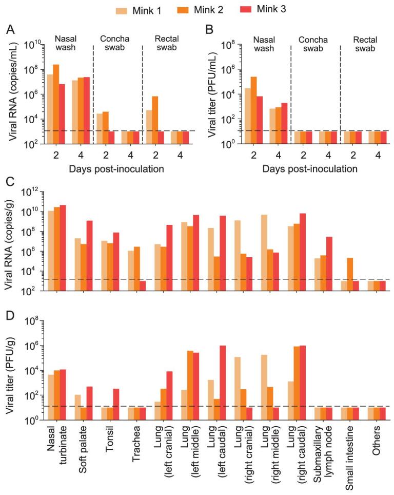
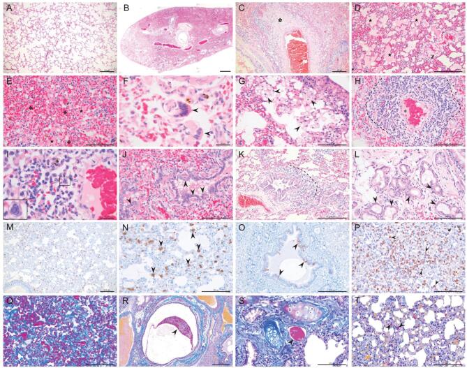
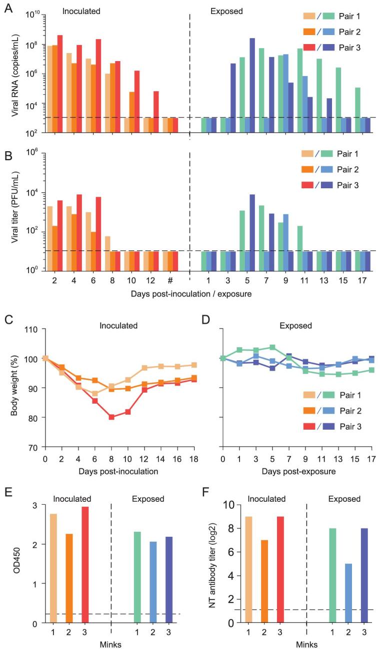
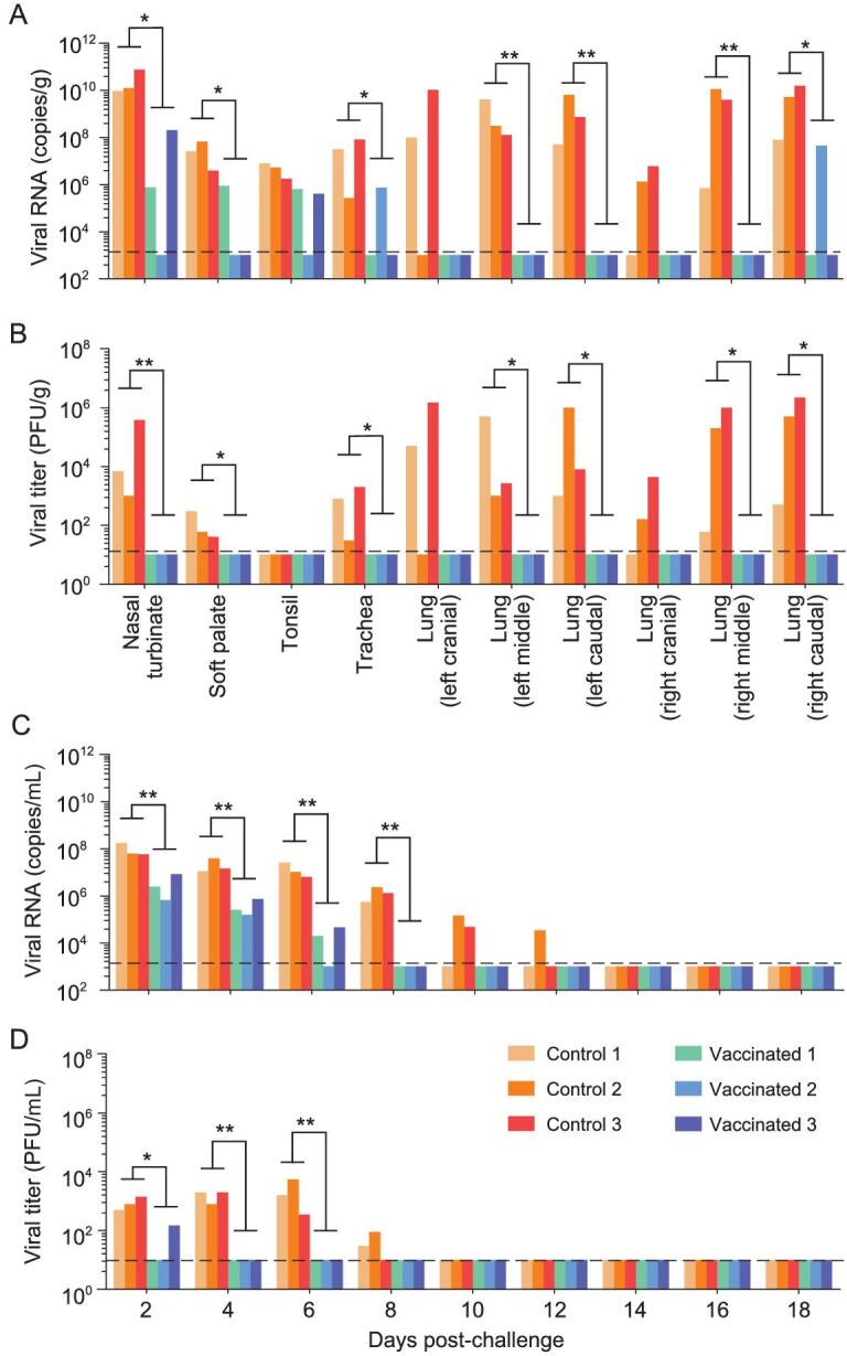
Minks are raised in many countries and have transmitted severe acute respiratory syndrome coronavirus 2 (SARS-CoV-2) to humans. However, the biologic properties of SARS-CoV-2 in minks are largely unknown. Here, we investigated and found that SARS-CoV-2 replicates efficiently in both the upper and lower respiratory tracts, and transmits efficiently in minks via respiratory droplets; pulmonary lesions caused by SARS-CoV-2 in minks are similar to those seen in humans with COVID-19. We further found that a spike protein-based subunit vaccine largely prevented SARS-CoV-2 replication and lung damage caused by SARS-CoV-2 infection in minks. Our study indicates that minks are a useful animal model for evaluating the efficacy of drugs or vaccines against COVID-19 and that vaccination is a potential strategy to prevent minks from transmitting SARS-CoV-2.

Keywords: SARS-CoV-2; animal model; mink; pathogenicity; replication; transmission.


Figures

Figure 1.
Figure 1.
Replication of SARS-CoV-2 in minks. Three minks were inoculated with HRB25 virus, and their nasal washes, concha swabs and rectal swabs were collected on days 2 and 4 post-infection (p.i.); organs were collected on day 4 p.i. for viral RNA detection and virus titration. (A) Viral RNA and (B) viral titers of nasal washes, concha swabs and rectal swabs of minks on days 2 and 4 p.i. (C) Viral RNA and (D) viral titers of organs of minks on day 4 p.i. ‘Others’ represents viral-negative organs, including the heart, kidneys, spleen, liver, pancreas and brain. Each color bar represents the value from an individual animal. The dashed lines indicate the lower limit of detection.
Figure 2.
Figure 2.
Histopathologic, immunohistochemical and histochemical studies. (A) Lung from control mink shows normal structure. (B-T) Lungs of minks infected with SARS-CoV-2. (B) Extensive and diffuse consolidation at sub-gross level. (C) Fibrinous necrosis of blood vessel wall (cinquefoil). (D) Intra-alveolar serous and fibrin exudation (asterisk). (E) Intra- and inter-alveolar hemorrhages (cloverleaf) and serous-fibrin exudation (asterisk). (F) Multinucleated syncytial cell in alveolar space (arrow). (G) Intra- and inter-alveolar foamy macrophage proliferation (arrow). (H) Severe lymphoplasmacytic perivasculitis and vasculitis (dotted line), and fibrin formation (asterisk) in a blood vessel. (I) Eosinophilic intranuclear inclusion body formation in a monocyte-like cell (■). (J) Degeneration of bronchiolar epithelium (arrow). (K) Peribronchiolar inflammatory infiltrate (dotted line). (L) Degeneration of bronchial gland epithelium. (M, N) Viral antigen-positive cells in the lung (arrow) and in (O) bronchi epithelium (arrow). (P) Marked proliferation of macrophages, monocytes and neutrophils, confirmed by immunohistochemistry with an anti-MAC387 antibody (arrow). (Q) Deposition of intra- and inter-alveolar collagen confirmed by Masson trichrome (blue) staining. (R) Embolus of mucous-fibrin mixture in bronchial lumen confirmed by Martius Scarlet Blue (MSB) staining (arrow). (S) Fibrin formation in a blood vessel confirmed by MSB staining (arrow). (T) Formation of microvascular thrombi confirmed by MSB staining (arrow). Scale bar in A, C, D, K, M and O = 200 μm; in E, G, H, J, L, N, P, Q, S and T = 100 μm; in F and I = 20 μm; and in R = 500 μm.
Figure 3.
Figure 3.
Transmission of SARS-CoV-2 in minks. Respiratory droplet transmission of SARS-CoV-2 HRB25 was evaluated in three pairs of minks. (A) Viral RNA in nasal washes of minks. (B) Viral titers in nasal washes of minks. (C) Body weight change of inoculated minks. (D) Body weight change of exposed minks. Antibodies against SARS-CoV-2 were detected by ELISA (E) and a neutralization assay (F). Hash represents the time points of days 14, 16 and 18 post-inoculation. Each color bar represents the value from an individual animal. The horizontal dashed lines indicate the lower limit of detection. OD450, optical density measured at 450 nm; NT antibody, neutralizing antibody.
Figure 4.
Figure 4.
Protective efficacy of a spike-based subunit vaccine against SARS-CoV-2 challenge in minks. Six vaccinated or control minks were challenged with SARS-CoV-2 HRB25. Three minks from each group were euthanized on day 4 post-challenge (p.c.) and their organs were collected for viral RNA detection (A) and infectious virus titration (B); nasal washes of the three other remaining minks in each group were collected for viral RNA detection (C) and infectious virus titration (D). The horizontal dashed lines indicate the lower limit of detection. Viral RNA and viral titers of control and vaccinated minks were statistically analyzed by using the one-tailed unpaired t-test. Single asterisk indicates P < 0.05, and double asterisks indicate P < 0.01.

Similar articles

Cited by

References

    1. Peiris JS, Lai ST, Poon LLet al. . Coronavirus as a possible cause of severe acute respiratory syndrome. Lancet 2003; 361: 1319–25.10.1016/S0140-6736(03)13077-2 - DOI - PMC - PubMed
    1. Zaki AM, van Boheemen S, Bestebroer TMet al. . Isolation of a novel coronavirus from a man with pneumonia in Saudi Arabia. N Engl J Med 2012; 367: 1814–20.10.1056/NEJMoa1211721 - DOI - PubMed
    1. Chan JF, Yuan S, Kok KHet al. . A familial cluster of pneumonia associated with the 2019 novel coronavirus indicating person-to-person transmission: a study of a family cluster. Lancet 2020; 395: 514–23.10.1016/S0140-6736(20)30154-9 - DOI - PMC - PubMed
    1. Zhu N, Zhang D, Wang Wet al. . A novel coronavirus from patients with pneumonia in China, 2019. N Engl J Med 2020; 382: 727–33.10.1056/NEJMoa2001017 - DOI - PMC - PubMed
    1. Mahase E. Coronavirus covid-19 has killed more people than SARS and MERS combined, despite lower case fatality rate. BMJ 2020; 368: m641.10.1136/bmj.m641 - DOI - PubMed