

Clinical applications of artificial intelligence and radiomics in neuro-oncology imaging
- PMID: 34676470
- PMCID: PMC8531173
- DOI: 10.1186/s13244-021-01102-6
Clinical applications of artificial intelligence and radiomics in neuro-oncology imaging
Abstract
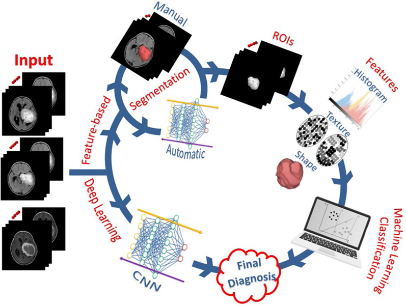
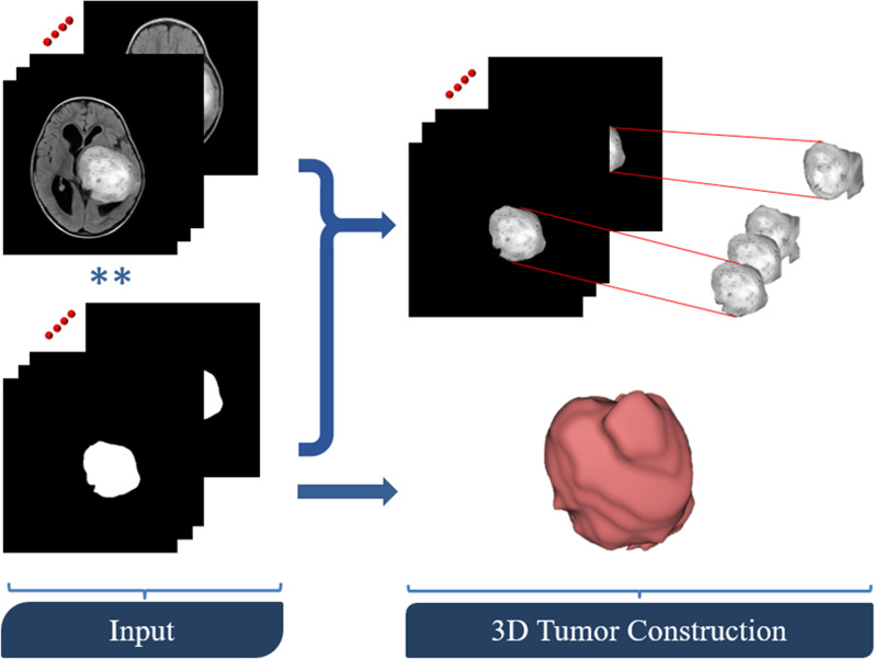
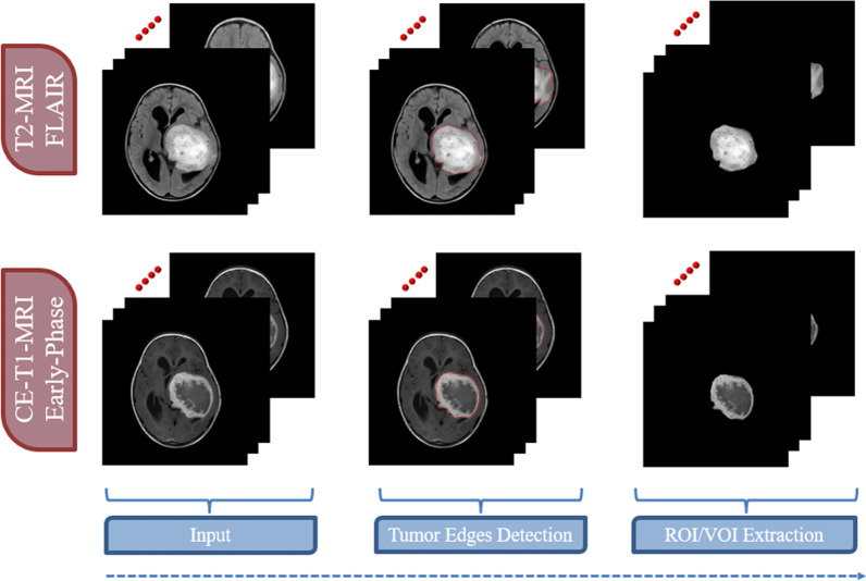
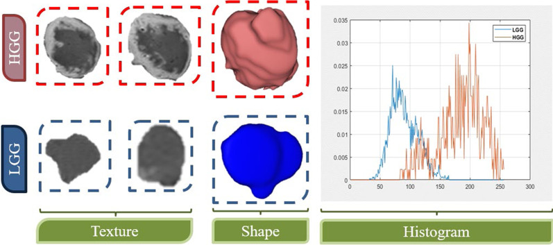
This article is a comprehensive review of the basic background, technique, and clinical applications of artificial intelligence (AI) and radiomics in the field of neuro-oncology. A variety of AI and radiomics utilized conventional and advanced techniques to differentiate brain tumors from non-neoplastic lesions such as inflammatory and demyelinating brain lesions. It is used in the diagnosis of gliomas and discrimination of gliomas from lymphomas and metastasis. Also, semiautomated and automated tumor segmentation has been developed for radiotherapy planning and follow-up. It has a role in the grading, prediction of treatment response, and prognosis of gliomas. Radiogenomics allowed the connection of the imaging phenotype of the tumor to its molecular environment. In addition, AI is applied for the assessment of extra-axial brain tumors and pediatric tumors with high performance in tumor detection, classification, and stratification of patient's prognoses.
Keywords: Artificial intelligence; Deep learning; Glioma; Machine learning; Radiomics.
© 2021. The Author(s).
Conflict of interest statement
The authors declare no competing interest.
Figures
References
Publication types
LinkOut - more resources
Full Text Sources
