. 2021 Nov 8;7(11):5028-5038.
doi: 10.1021/acsbiomaterials.1c00817. Epub 2021 Oct 22.

Soft Hydrogel Inspired by Elastomeric Proteins

Affiliations

Soft Hydrogel Inspired by Elastomeric Proteins

Antonietta Pepe et al. ACS Biomater Sci Eng. .

Abstract

Elastin polypeptides based on -VPGVG- repeated motifs are widely used in the production of biomaterials because they are stimuli-responsive systems. On the other hand, glycine-rich sequences, mainly present in tropoelastin terminal domains, are responsible for the elastin self-assembly. In a previous study, we have recombinantly expressed a chimeric polypeptide, named resilin, elastin, and collagen (REC), inspired by glycine-rich motifs of elastin and containing resilin and collagen sequences as well. Herein, a three-block polypeptide, named (REC)3, was expressed starting from the previous monomer gene by introducing key modifications in the sequence. The choice was mandatory because the uneven distribution of the cross-linking sites in the monomer precluded the hydrogel production. In this work, the cross-linked polypeptide appeared as a soft hydrogel, as assessed by rheology, and the linear un-cross-linked trimer self-aggregated more rapidly than the REC monomer. The absence of cell-adhesive sequences did not affect cell viability, while it was functional to the production of a material presenting antiadhesive properties useful in the integration of synthetic devices in the body and preventing the invasion of cells.

Keywords: antiadhesive materials; circular dichroism; cytocompatibility; elastin; hydrogel.


Conflict of interest statement

The authors declare no competing financial interest.

Figures

Figure 1
Figure 1
Schematic representation of the primary structure of the (REC)3 polypeptide.
Figure 2
Figure 2
12% SDS-PAGE analysis of (REC)3 production screening (A) and purification process (B). (A) Total protein fraction analysis of E. coli BLR(DE3) after overnight induction in a modified TB medium. Lane M: protein marker; lane Ø—negative control, untransformed BLR(DE3); and Lanes 1–5, five transformed colonies were randomly selected and analyzed. The red marker highlights the (REC)3 polypeptide production by different transformants. (B) Increasing amounts of the purified (REC)3 polypeptide were evaluated: 5 μg (lane 1), 10 μg (lane 2), and 15 μg (lane 3); M: protein marker, the molecular weight of bands is indicated.
Figure 3
Figure 3
(a) CD spectra of the (REC)3 polypeptide in PBS (filled symbols) and TFE (open symbols) at the indicated temperatures: 0 °C (squares); 25 °C (circles); 37 °C (diamonds); and 60 °C (triangles); (b) CD spectra of the REC polypeptide in an aqueous solution (filled symbols) and TFE (open symbols) at the indicated temperatures: 0 °C (squares); 25 °C (circles); and 60 °C (triangles).
Figure 4
Figure 4
TEM micrographs of (REC)3 incubated at 37 °C after drop deposition (a) and after 24 h (b).
Figure 5
Figure 5
AFM images of (REC)3 incubated at 37 °C after 0 (a) and 24 (b) h.
Figure 6
Figure 6
(a) Hydrogel; (b) SEM image of the hydrogel, where bar represents 30 μm; (c) evolution of G′ and G″ with frequency at 1% strain: average storage modulus (black) and loss modulus (red); and (d) dependence of the complex modulus magnitude with f1/2.
Figure 7
Figure 7
Viability percentage as the metabolic activity of HUVECs and MSCs with respect to untreated cells in the same conditions. Cells were incubated with 5 mg/mL (REC)3 for 24 or 72 h. Metabolic active cell numbers were measured, each sample four-fold, using the Alamar Blue assay kit in three independent experiments. No statistically significant differences are observed. The error bars represent the standard deviation.
Figure 8
Figure 8
Viability of HUVECs and MSCs cultured on surfaces coated with (REC)3 solutions at 5 or 10 mg/mL concentrations. Histogram represents the percentages of live cells after 4 days of incubation with respect to live cells cultured on a standard tissue culture support. The error bars correspond to the standard deviation.
Figure 9
Figure 9
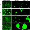
Panels A (10×) and B (20×): representative images of HUVECs cultured on fibronectin (positive control), 5 mg/mL or 10 mg/mL (REC)3-coated surfaces. Panels C (10×) and D (20×): representative images of hMSCs cultured on fibronectin (positive control), 5 mg/mL or 10 mg/mL (REC)3-coated surfaces. The living cells fluoresce green, whereas dead cells with compromised membranes fluoresce red.

References

    1. Huettner N.; Dargaville T. R.; Forget A. Discovering Cell-Adhesion Peptides in Tissue Engineering: Beyond RGD. Trends Biotechnol. 2018, 36, 372–383. 10.1016/j.tibtech.2018.01.008. - DOI - PubMed
    1. Werkmeister J. A.; Ramshaw J. A. M. Recombinant protein scaffolds for tissue engineering. Biomed. Mater. 2012, 7, 012002.10.1088/1748-6041/7/1/012002. - DOI - PubMed
    1. Gosline J.; Lillie M.; Carrington E.; Guerette P.; Ortlepp C.; Savage K. Elastic proteins: biological roles and mechanical properties. Philos. Trans. R. Soc., B 2002, 357, 121–132. 10.1098/rstb.2001.1022. - DOI - PMC - PubMed
    1. Su R. S.-C.; Kim Y.; Liu J. C. Resilin: protein-based elastomeric biomaterials. Acta Biomater. 2014, 10, 1601–1611. 10.1016/j.actbio.2013.06.038. - DOI - PubMed
    1. Elvin C. M.; Carr A. G.; Huson M. G.; Maxwell J. M.; Pearson R. D.; Vuocolo T.; Liyou N. E.; Wong D. C. C.; Merritt D. J.; Dixon N. E. Synthesis and properties of crosslinked recombinant pro-resilin. Nature 2005, 437, 999–1002. 10.1038/nature04085. - DOI - PubMed

Publication types