. 2021 Dec;24(6):865.
doi: 10.3892/mmr.2021.12505. Epub 2021 Oct 22.

MicroRNA‑200c‑3p suppresses intervertebral disc degeneration by targeting RAP2C/ERK signaling

Affiliations

MicroRNA‑200c‑3p suppresses intervertebral disc degeneration by targeting RAP2C/ERK signaling

Jianping Cao et al. Mol Med Rep. 2021 Dec.

Abstract

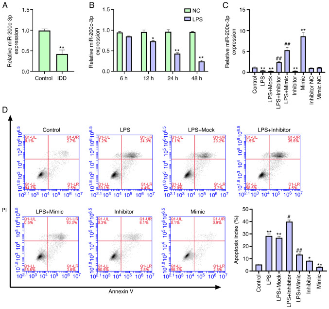
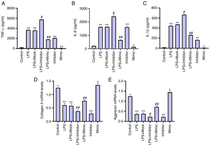
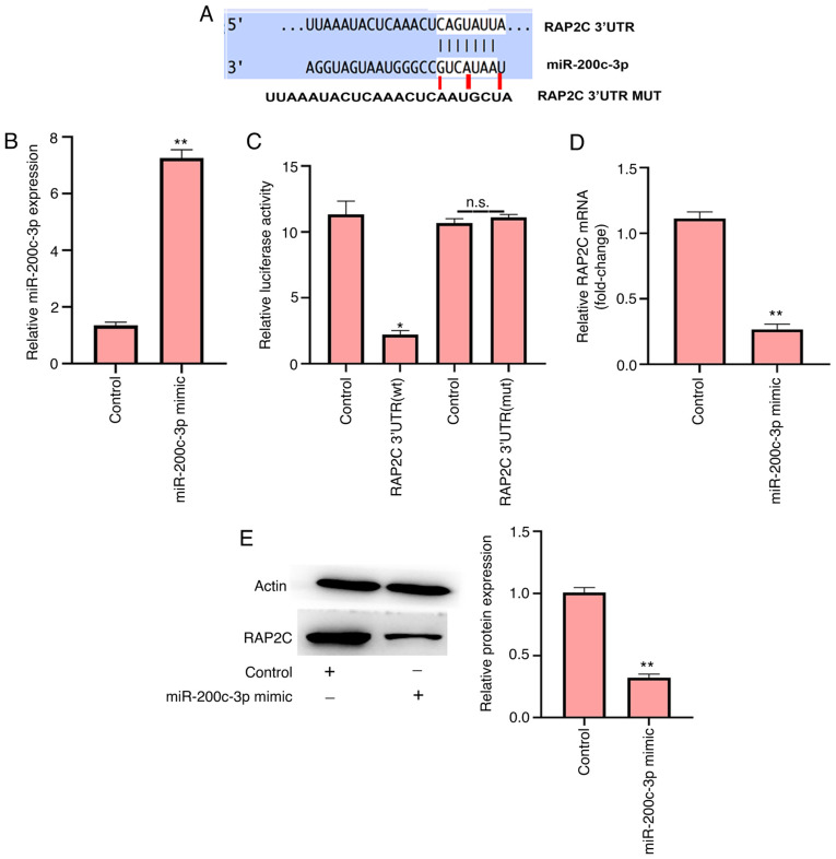
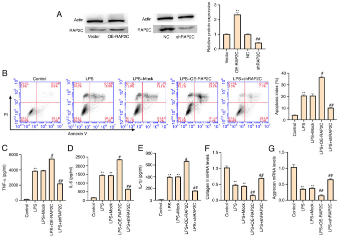
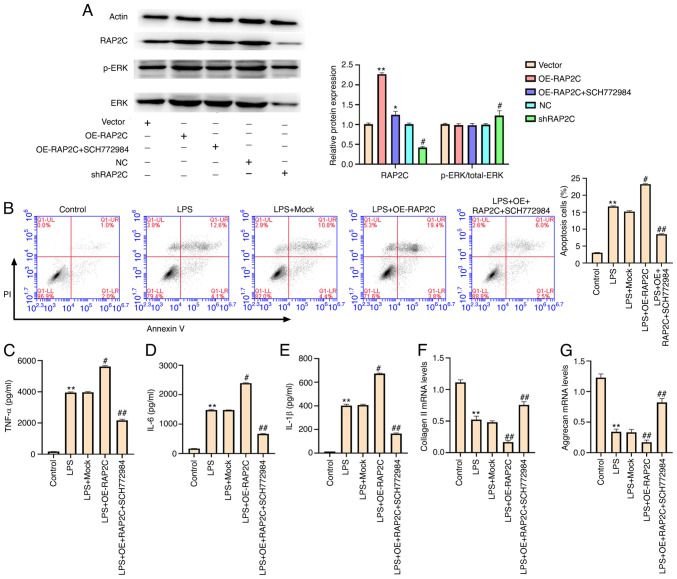
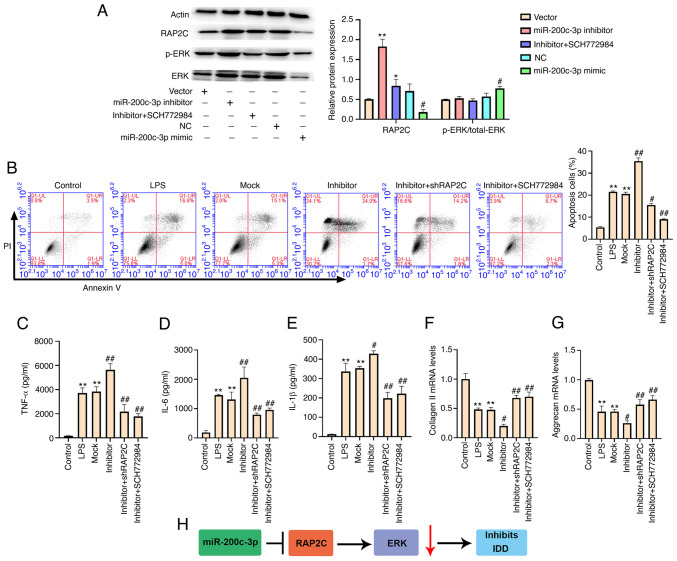
Intervertebral disc degeneration (IDD) is a major cause of lower back pain. The high morbidity associated with this disease diminishes the quality of life of those who are affected. MicroRNAs (miRs) play crucial roles in various diseases, including IDD. However, the mechanism via which miR‑200c‑3p plays a role in the development of IDD remains unknown. The present study aimed to investigate the effect of miR‑200c‑3p on the progression of IDD and the underlying mechanism. The expression level of miR‑200c‑3p was evaluated in intervertebral disc tissues from patients with IDD. To construct the IDD cell model, the nucleus pulposus (NP) cells were treated with lipopolysaccharide (LPS) 24 h following transfection with miR‑200c‑3p mimic or inhibitor. A luciferase activity assay was performed, while reverse transcription‑quantitative PCR and western blotting were conducted to determine the RNA and protein expression levels, respectively. The expression level of miR‑200c‑3p in the intervertebral disc tissues of patients with IDD was lower than that of normal subjects. LPS treatment reduced the expression level of miR‑200c‑3p in NP cells. Moreover, miR‑200c‑3p mimic inhibited LPS‑induced NP cell apoptosis. It was found that miR‑200c‑3p attenuated inflammatory cytokine levels and extracellular matrix (ECM) degradation in NP cells. Furthermore, miR‑200c‑3p targeted Ras‑related protein 2C (RAP2C) in NP cells. RAP2C promoted apoptosis, inflammatory cytokine levels and ECM degradation by activating ERK signaling. Knockdown of RAP2C and inhibition of ERK signaling by SCH772984 partially reversed the proinflammatory effect of the miR‑200c‑3p inhibitor on LPS‑treated NP cells. Thus, miR‑200c‑3p inhibits NP cell apoptosis, inflammatory cytokine levels and ECM degradation in IDD by targeting RAP2C/ERK signaling.

Keywords: ERK signaling; IDD; NP cells; RAP2C; miR‑200c‑3p.


Conflict of interest statement

The authors declare that they have competing interests.

Figures

Figure 1.
Figure 1.
miR-200c-3p inhibits LPS-induced NP cell apoptosis. (A) Expression levels of miR-200c-3p were measured via RT-qPCR in the intervertebral disc tissues of patients with IDD (n=22) and normal cases (n=9). (B) Expression levels of miR-200c-3p were assessed via RT-qPCR in NP cells treated with LPS (1 µg/ml). *P<0.05, **P<0.01 vs. NC. NP cells were treated with LPS (1 µg/ml) or co-treated with LPS (1 µg/ml) and miR-200c-3p mimic, inhibitor or corresponding control. (C) Expression levels of miR-200c-3p were analyzed via RT-qPCR. (D) Cell apoptosis was tested using flow cytometry analysis. Data are presented as the mean ± SD. *P<0.05, **P<0.01 vs. control; #P<0.05, ##P<0.01 vs. LPS. LPS, lipopolysaccharide; RT-qPCR, reverse transcription-quantitative PCR; miR, microRNA; NC, negative control; IDD, intervertebral disc degeneration; NP, nucleus pulposus.
Figure 2.
Figure 2.
miR-200c-3p attenuates inflammatory cytokine levels and extracellular matrix degradation in LPS-treated NP cells. NP cells were treated with LPS (1 µg/ml) or co-treated with LPS (1 µg/ml) and miR-200c-3p mimic, inhibitor. Levels of (A) TNF-α, (B) IL-6 and (C) IL-1β in the culture medium of the cells were analyzed using ELISAs. Expression levels of (D) collagen II and (E) aggrecan were examined via reverse transcription-quantitative PCR. Data are presented as the mean ± SD. *P<0.05, **P<0.01 vs. control; #P<0.05, ##P<0.01 vs. LPS. LPS. LPS, lipopolysaccharide; miR, microRNA; NP, nucleus pulposus.
Figure 3.
Figure 3.
miR-200c-3p targets RAP2C in nucleus pulposus cells. (A) Interaction of miR-200c-3p and RAP2C 3′UTR was identified via bioinformatics analysis using TargetScan. (B) NP cells were transfected with control mimic or miR-200c-3p mimic. (C) Luciferase activities of RAP2C WT and RAP2C with the miR-200c-3p-binding site mutant (RAP2C MUT) were determined using luciferase reporter gene assays. (D) mRNA expression level of RAP2C was assessed using reverse transcription-quantitative PCR in the cells. (E) Protein expression level of RAP2C was measured via western blotting. Data are presented as the mean ± SD. *P<0.05, **P<0.01 vs. control. ns, no significance; miR, microRNA; WT, wild-type; MUT, mutant; RAP2C, Ras-related protein 2C; UTR, untranslated region.
Figure 4.
Figure 4.
RAP2C promotes apoptosis, inflammatory cytokine levels and ECM degradation in LPS-treated NP cells. LPS (1 µg/ml)-treated NP cells were infected with lentiviral plasmids carrying RAP2C shRNA or the corresponding control shRNA, or transfected with the control vector or the vector carrying the complete RAP2C coding sequence for overexpression. (A) Expression levels of RAP2C were measured via western blotting. **P<0.01 vs. vector; ##P<0.01 vs. NC. (B) Cell apoptosis was measured using flow cytometry analysis. Levels of (C) TNF-α, (D) IL-6 and (E) IL-1β in the culture medium of the cells were analyzed using ELISAs. Expression levels of (F) collagen II and (G) aggrecan were examined via reverse transcription-quantitative PCR. Data are presented as the mean ± SD. **P<0.01 vs. control; #P<0.05, ##P<0.01 vs. LPS. LPS, lipopolysaccharide; shRNA/sh, short hairpin RNA; NC, negative control; RAP2C, Ras-related protein 2C; NP, nucleus pulposus; OE, overexpression vector.
Figure 5.
Figure 5.
RAP2C enhances apoptosis, inflammatory cytokine levels and extracellular matrix degradation by activating ERK signaling. (A) NP cells were infected with the lentiviral plasmids carrying RAP2C shRNA or the corresponding control shRNA, transfected with the control vector or the vector carrying the complete RAP2C coding sequence for overexpression or co-treated with the RAP2C overexpression vector and the ERK inhibitor SCH772984 (10 µmol/l). The expression levels of RAP2C, ERK and β-actin, and the phosphorylation of ERK (p-ERK) were examined via western blotting. *P<0.05, **P<0.01 vs. vector; #P<0.05 vs. NC. LPS (1 µg/ml)-treated NP cells were transfected with the control vector, or the vector carrying the complete RAP2C coding sequence for overexpression or co-treated with RAP2C overexpression vector and the ERK inhibitor SCH772984 (10 µmol/l). (B) Cell apoptosis was examined via flow cytometry analysis. Levels of (C) TNF-α, (D) IL-6 and (E) IL-1β in the culture medium of the cells were tested using ELISAs. Expression levels of (F) collagen II and (G) aggrecan were determined using reverse transcription-quantitative PCR. Data are presented as the mean ± SD. *P<0.05, **P<0.01 vs. control; #P<0.05, ##P<0.01 vs. LPS. LPS, lipopolysaccharide; shRNA/sh, short hairpin RNA; NC, negative control; RAP2C, Ras-related protein 2C; NP, nucleus pulposus; OE, overexpression vector; p-, phosphorylated.
Figure 6.
Figure 6.
miR-200c-3p suppresses IDD by targeting RAP2C/ERK signaling. (A) NP cells were transfected with miR-200c-3p mimic, inhibitor or corresponding control, or co-treated with miR-200c-3p inhibitor and the ERK inhibitor SCH772984 (10 µmol/l). The expression levels of RAP2C, ERK and β-actin, and the phosphorylation of ERK (p-ERK) were examined via western blotting. LPS (1 µg/ml)-treated NP cells were transfected with miRNA control inhibitor or miR-200c-3p inhibitor, or co-transfected with miR-200c-3p inhibitor and the lentiviral plasmids carrying RAP2C shRNA, or co-transfected with miR-200c-3p inhibitor and the ERK inhibitor SCH772984 (10 µmol/l). *P<0.05, **P<0.01 vs. vector; #P<0.05 vs. NC. (B) Cell apoptosis was analyzed using flow cytometry. The levels of (C) TNF-α, (D) IL-6 and (E) IL-1β in the culture medium of the cells were assessed using ELISA. The expression levels of (F) collagen II and (G) aggrecan were examined using reverse transcription-quantitative PCR. (H) The regulatory pathway of miR-200c-3p in LPS-treated NP cells. Data are presented as the mean ± SD. **P<0.01 vs. control; #P<0.05, ##P<0.01 vs. LPS. IDD, intervertebral disc degeneration; LPS, lipopolysaccharide; shRNA/sh, short hairpin RNA; NC, negative control; RAP2C, Ras-related protein 2C; NP, nucleus pulposus; p-, phosphorylated; miRNA/miR, microRNA.

Similar articles

Cited by

References

    1. Wang SZ, Chang Q, Lu J, Wang C. Growth factors and platelet-rich plasma: Promising biological strategies for early intervertebral disc degeneration. Int Orthop. 2015;39:927–934. doi: 10.1007/s00264-014-2664-8. - DOI - PubMed
    1. Tisherman R, Coelho P, Phillibert D, Wang D, Dong Q, Vo N, Kang J, Sowa G. NF-κB signaling pathway in controlling intervertebral disk cell response to inflammatory and mechanical stressors. Phys Ther. 2016;96:704–711. doi: 10.2522/ptj.20150045. - DOI - PMC - PubMed
    1. Zhang Y, Yang J, Zhou X, Wang N, Li Z, Zhou Y, Feng J, Shen D, Zhao W. Knockdown of miR-222 inhibits inflammation and the apoptosis of LPS-stimulated human intervertebral disc nucleus pulposus cells. Int J Mol Med. 2019;44:1357–1365. - PMC - PubMed
    1. Zhou Q, Huang SX, Zhang F, Li SJ, Liu C, Xi YY, Wang L, Wang X, He QQ, Sun CC, Li DJ. MicroRNAs: A novel potential biomarker for diagnosis and therapy in patients with non-small cell lung cancer. Cell Prolif. 2017;50:e12394. doi: 10.1111/cpr.12394. - DOI - PMC - PubMed
    1. Zhang Q, Weng Y, Jiang Y, Zhao S, Zhou D, Xu N. Overexpression of miR-140-5p inhibits lipopolysaccharide-induced human intervertebral disc inflammation and degeneration by downregulating toll-like receptor 4. Oncol Rep. 2018;40:793–802. - PubMed