. 2021 Dec;9(6):e00822.
doi: 10.1002/prp2.822.

Chemogenetic stimulation of the Gi pathway in astrocytes suppresses neuroinflammation

Affiliations

Chemogenetic stimulation of the Gi pathway in astrocytes suppresses neuroinflammation

Jae-Hong Kim et al. Pharmacol Res Perspect. 2021 Dec.

Abstract

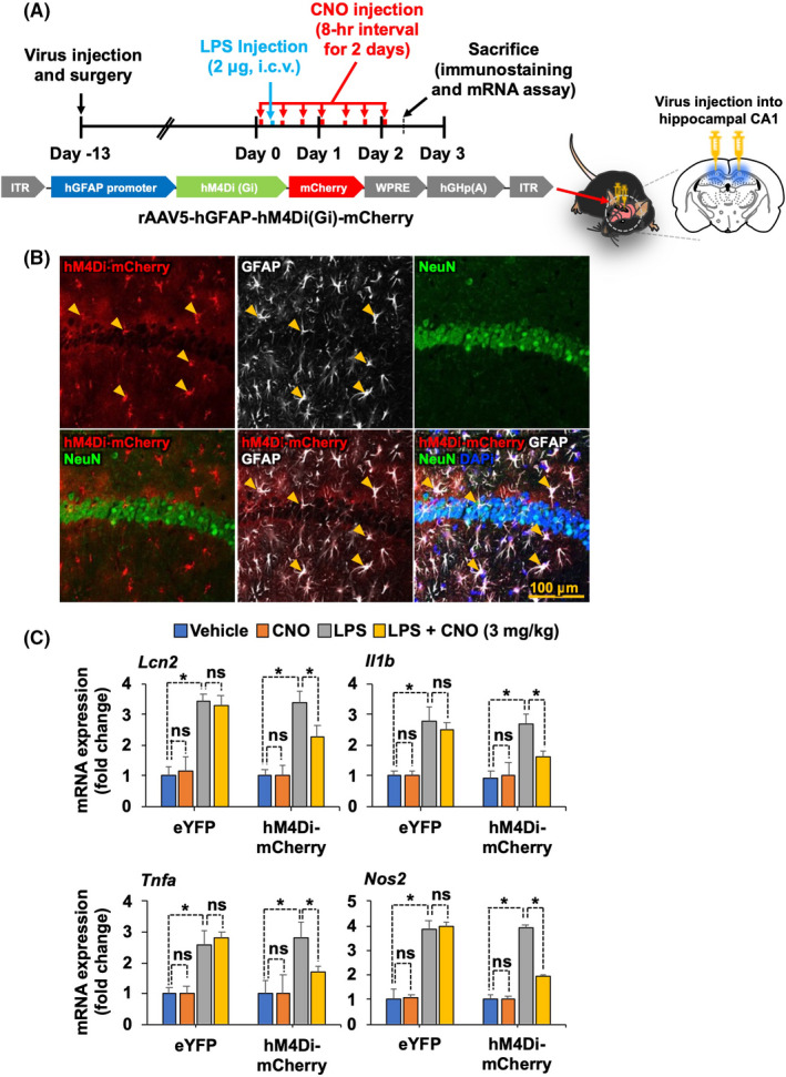
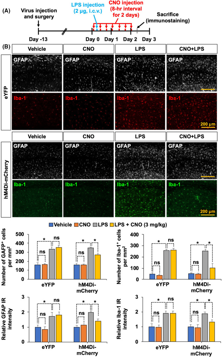
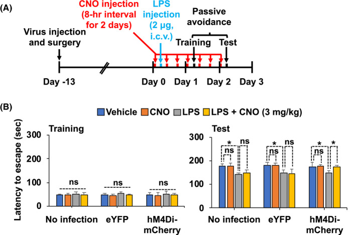
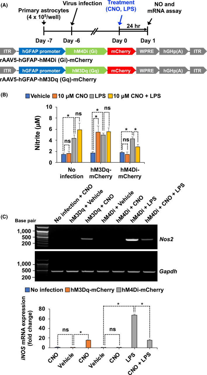
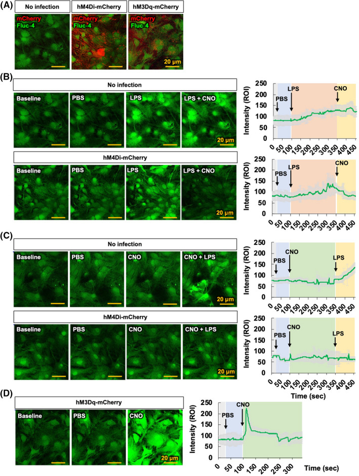
Engineered G protein-coupled receptors (GPCRs) are commonly used in chemogenetics as designer receptors exclusively activated by designer drugs (DREADDs). Although several GPCRs have been studied in astrocytes using a chemogenetic approach, the functional role of the astrocytic Gi pathway is not clear, as the literature is conflicting depending on the brain regions or behaviors investigated. In this study, we evaluated the role of the astrocytic Gi pathway in neuroinflammation using a Gi -coupled DREADD (hM4Di). Gi -DREADD was expressed in hippocampal astrocytes of a lipopolysaccharide (LPS)-induced neuroinflammation mouse model using adeno-associated viruses. We found that astrocyte Gi -DREADD stimulation using clozapine N-oxide (CNO) inhibits neuroinflammation, as characterized by decreased levels of proinflammatory cytokines, glial activation, and cognitive impairment in mice. Subsequent experiments using primary astrocyte cultures revealed that Gi -DREADD stimulation significantly downregulated LPS-induced expression of Nos2 mRNA and nitric oxide production. Similarly, in vitro calcium imaging showed that activation of the astrocytic Gi pathway attenuated intracellular calcium transients triggered by LPS treatment, suggesting a positive correlation between enhanced calcium transients and the inflammatory phenotype of astrocytes observed in the inflamed brain. Taken together, our results indicate that the astrocytic Gi pathway plays an inhibitory role in neuroinflammation, providing an opportunity to identify potential cellular and molecular targets to control neuroinflammation.

Keywords: Gi-DREADD; astrocyte; chemogenetics; hM4Di; neuroinflammation.


Conflict of interest statement

The authors declare no conflicts of interest in regards to this manuscript.

Figures

FIGURE 1
FIGURE 1
Selective expression and activation of astrocytic hM4Di inhibit LPS‐induced expression of proinflammatory mediators in the mouse hippocampus. (A) Schematic diagram showing the timeline of experimentation and AAV vector constructs. (B) Confocal images of mCherry labeling and immunofluorescence analysis. Astrocytes were immunohistochemically labeled with GFAP (white), and neurons were labeled with NeuN (green). Nuclei were stained with DAPI (blue). Arrowheads indicate the colocalization of hGFAP‐hM4Di‐mCherry and cell‐type‐specific markers. Scale bar, 200 μm. (C) After behavior tests, mice were sacrificed and total mRNA was extracted from the hippocampal tissues of each group. RT‐PCR was performed to assess the expression levels of Lcn2, Il1b, Tnfa, and Nos2 mRNA expression profiles are displayed as the fold increase of gene expression normalized to Gapdh mRNA levels. Results are expressed as means ± SEM (n = 4). *p < .05 between the indicated groups (Student's t‐test). eYFP, AAV5‐hGFAP‐eYFP; hM4Di‐mCherry, AAV5‐hGFAP‐hM4Di‐mCherry; ns, not significant
FIGURE 2
FIGURE 2
Selective activation of astrocytic hM4Di inhibits LPS‐induced glial activation in the mouse hippocampus. (A) Schematic diagram showing the timeline of experimentation. (B) Immunofluorescence staining and image analysis were performed. Astrocytes were immunohistochemically labeled with GFAP (white), and microglia were labeled with Iba‐1 (red or green). Quantitative analysis of GFAP‐positive astrocytes and Iba‐1‐positive microglia as well as relative GFAP and Iba‐1 immunoreactivity (IR) intensity in the hippocampus are presented in adjacent graphs. Scale bar, 200 μm. Results are expressed as means ± SEM (n = 4). *p < .05 between the indicated groups (one‐way ANOVA with Bonferroni's post hoc test). eYFP, AAV5‐hGFAP‐eYFP; hM4Di‐mCherry, AAV5‐hGFAP‐hM4Di; IR, immunoreactivity; ns, not significant
FIGURE 3
FIGURE 3
Selective activation of astrocytic hM4Di alleviates LPS‐induced cognitive impairment. (A) Schematic diagram showing the timeline of experimentation. (B) Cognitive impairment was evaluated using the passive avoidance test. The results are expressed as means ± SEM (n = 5 for each group). *p < .05 between the indicated groups (one‐way ANOVA with Bonferroni's post hoc test). eYFP, AAV5‐hGFAP‐eYFP; hM4Di‐mCherry, AAV5‐hGFAP‐hM4Di‐mCherry; ns, not significant
FIGURE 4
FIGURE 4
Stimulation of astrocytic Gi signaling in culture decreases LPS‐induced levels of nitrite and expression of Nos2 mRNA. (A) Schematic diagram showing the timeline of experimentation and AAV vector constructs. (B) Primary astrocyte cultures were treated with CNO and LPS for 20 min. The levels of nitrite in culture media were measured after 24 h. (C) Total cellular RNA was extracted 24 h after treatment. Expression levels of Nos2 mRNA were determined by RT‐PCR. Data were normalized to mRNA levels of Gapdh, and results are expressed as means ± SEM (n = 6). *p < .05 between the indicated groups (Student's t‐test). eYFP, AAV5‐hGFAP‐eYFP; hM3Dq‐mCherry, AAV5‐hM3Dq‐mCherry; hM4Di‐mCherry, AAV5‐hM4Di‐mCherry; ns, not significant
FIGURE 5
FIGURE 5
Gi signaling activation in astrocytes inhibits LPS‐induced intracellular Ca2+ levels. (A) Representative images showing co‐localization of Fluo‐4‐AM (green) and mCherry (red) in negative controls, hM4Di‐mCherry‐, and hM3Dq‐mCherry‐infected primary astrocyte cultures. (B, C) Representative images showing Ca2+ transients after treatment with CNO (10 μM) in negative controls and hM4Di‐expressing primary astrocytes loaded with Fluo‐4‐AM. Astrocytes were treated with PBS at 20 s, LPS (100 ng/ml) at 100 s, and CNO at 350 s (B); or treated with PBS at 20 s, CNO at 100 s, and LPS (100 ng/ml) at 350 s (C). Representative traces show the change of intracellular Ca2+ in astrocytes as induced by LPS in the absence or presence of CNO. (D) hM3Dq‐expressing primary astrocytes loaded with Fluo‐4‐AM exhibited Ca2+ transients after treatment with CNO. Astrocytes were treated with PBS at 20 s and CNO at 100 s. Representative traces show the change of intracellular Ca2+ in astrocytes induced by CNO. Arrows indicate the treatment time for PBS, LPS, and CNO in the traces. Results are expressed as means ± SEM (n = 6). Scale bar, 20 μm. eYFP, AAV5‐hGFAP‐eYFP; hM3Dq‐mCherry, AAV5‐hM3Dq‐mCherry; hM4Di‐mCherry, AAV5‐hM4Di‐mCherry

Similar articles

Cited by

References

    1. Urban DJ, Roth BL. DREADDs (designer receptors exclusively activated by designer drugs): chemogenetic tools with therapeutic utility. Annu Rev Pharmacol Toxicol. 2015;55:399‐417. - PubMed
    1. Hirbec H, Déglon N, Foo LC, et al. Emerging technologies to study glial cells. Glia. 2020;68:1692‐1728. - PubMed
    1. Grace PM, Wang X, Strand KA, et al. DREADDed microglia in pain: implications for spinal inflammatory signaling in male rats. Exp Neurol. 2018;304:125‐131. - PMC - PubMed
    1. Pelvig DP, Pakkenberg H, Stark AK, Pakkenberg B. Neocortical glial cell numbers in human brains. Neurobiol Aging. 2008;29:1754‐1762. - PubMed
    1. Nam MH, Han KS, Lee J, et al. Activation of astrocytic mu‐opioid receptor causes conditioned place preference. Cell Rep. 2019;28:1154‐1166 e5. - PubMed

Publication types

MeSH terms

LinkOut - more resources