Review
. 2021 Nov 2;54(21):4012-4023.
doi: 10.1021/acs.accounts.1c00521. Epub 2021 Oct 22.

Immunogenicity of In Vitro-Transcribed RNA

Affiliations
Review

Immunogenicity of In Vitro-Transcribed RNA

Xin Mu et al. Acc Chem Res. .

Abstract

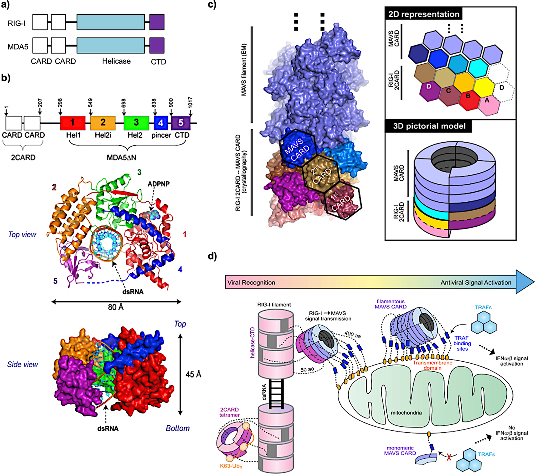
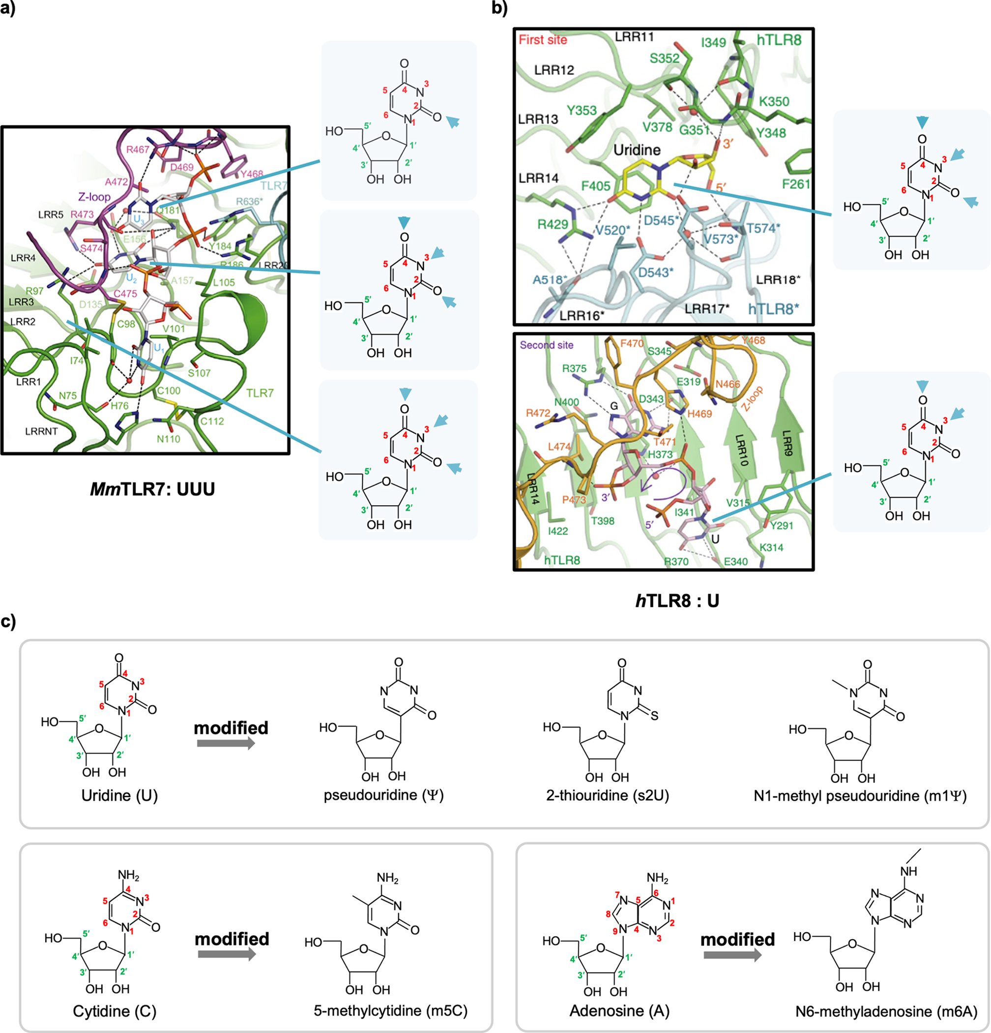
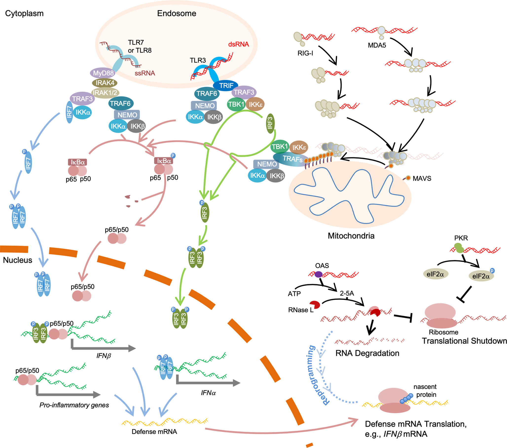
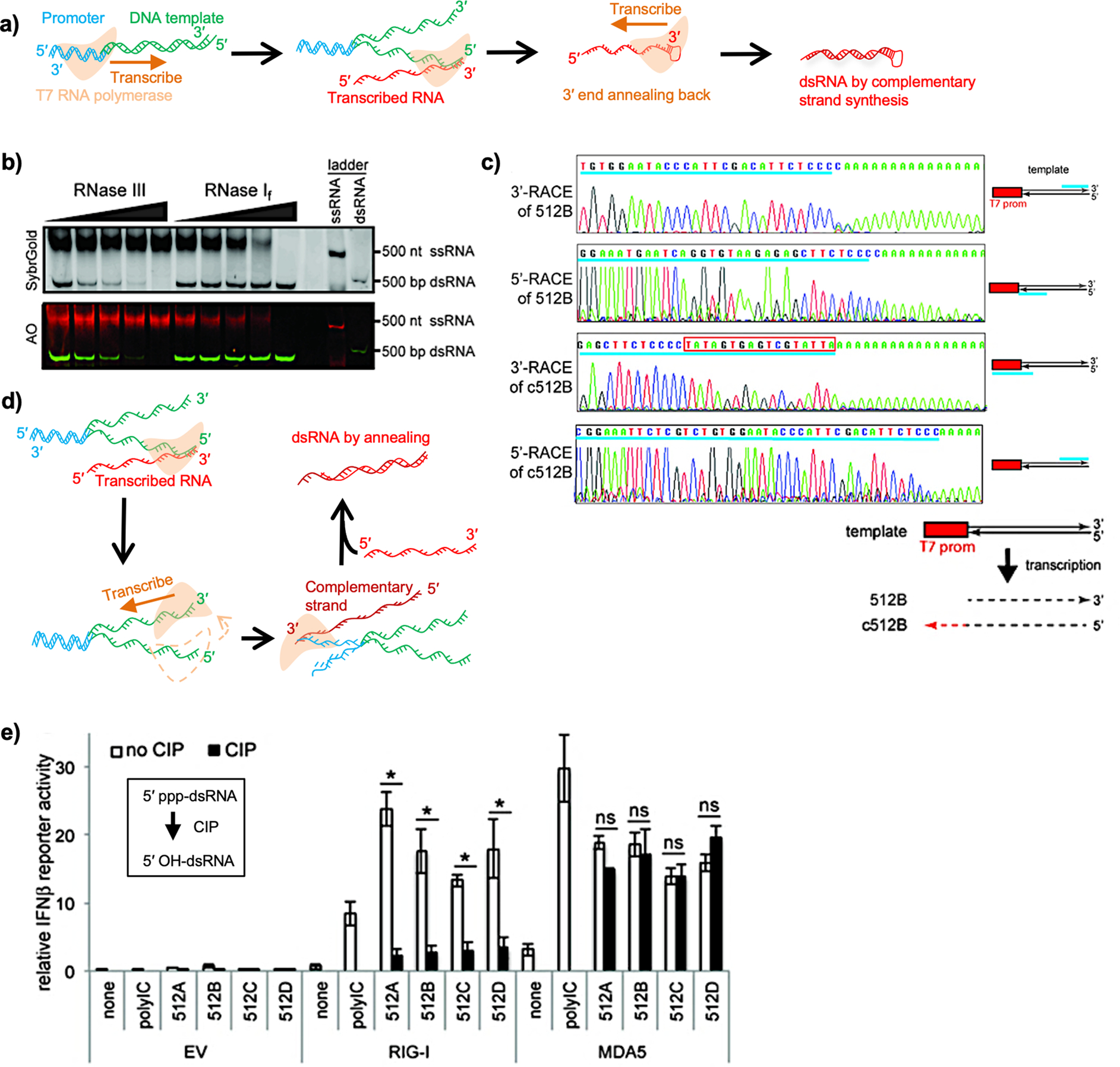
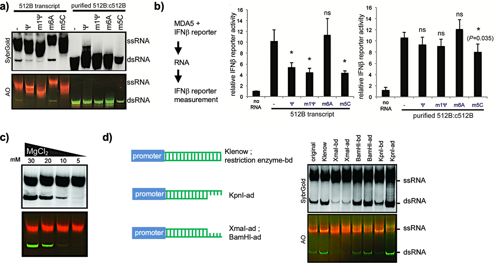
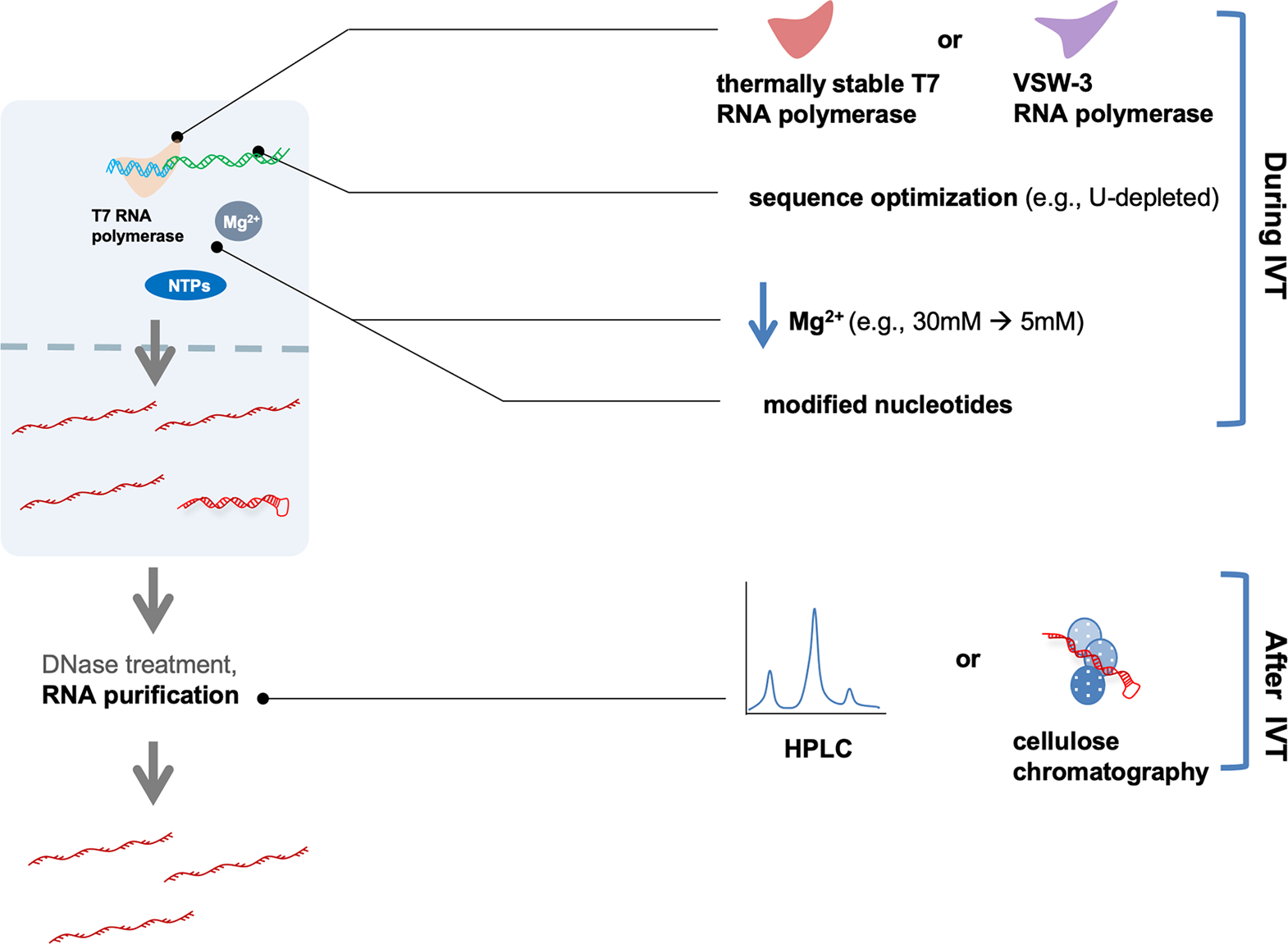
In vitro-transcribed RNAs are emerging as new biologics for therapeutic innovation, as exemplified by their application recently in SARS-CoV-2 vaccinations. RNAs prepared by in vitro transcription (IVT) allow transient expression of proteins of interest, conferring safety over DNA- or virus-mediated gene delivery systems. However, in vitro-transcribed RNAs should be used with caution because of their immunogenicity, which is in part triggered by double-stranded RNA (dsRNA) byproducts during IVT. Cellular innate immune response to dsRNA byproducts can lead to undesirable consequences, including suppression of protein synthesis and cell death, which in turn can detrimentally impact the efficacy of mRNA therapy. Thus, it is critical to understand the nature of IVT byproducts and the mechanisms by which they trigger innate immune responses.Our lab has been investigating the mechanisms by which the innate immune system discriminates between "self" and "nonself" RNA, with the focus on the cytoplasmic dsRNA receptors retinoic acid-inducible gene I (RIG-I) and melanoma differentiation-associated 5 (MDA5). We have biochemically and structurally characterized critical events involving RNA discrimination and signal transduction by RIG-I or MDA5. We have used in vitro-transcribed RNAs as tools to investigate RNA specificity of RIG-I and MDA5, which required optimization of the IVT reaction and purification processes to eliminate the effect of IVT byproducts. In this Account, we summarize our current understanding of RIG-I and MDA5 and IVT reactions and propose future directions for improving IVT as a method to generate both research tools and therapeutics. Other critical proteins in cellular innate immune response to dsRNAs are also discussed. We arrange the contents in the following order: (i) innate immunity sensors for nonself RNA, including the RIG-I-like receptors (RLRs) in the cytosol and the toll-like receptors (TLRs) in the endosome, as well as cytoplasmic dsRNA-responding proteins, including protein kinase R (PKR) and 2',5'-oligoadenylate synthetases (OASes), illustrating the feature of protein-RNA binding and its consequences; (ii) the immunogenicity of IVT byproducts, specifically the generation of dsRNA molecules during IVT; and (iii) methods to reduce IVT RNA immunogenicity, including optimizations of RNA polymerases, reagents, and experimental conditions during IVT and subsequent purification.


Conflict of interest statement

The authors declare no competing financial interest.

Figures

Figure 1.
Figure 1.
(a) Schematic of the domain organization of RIG-I and MDA5. (b) Top and side views of MDA5 (without 2CARDs): 12 bp dsRNA complex structure in the presence of the ADP analogue ADPNP. (c) Structures of MAVS CARD filament nucleated by RIG-I 2CARD oligomers. 2D and 3D models are shown on the right. (d) Cartoon model of filamentous RLRs nucleating MAVS to form filaments to activate downstream factors for IFN signaling. (b) Reprinted with permission from ref . Copyright 2013 Elsevier Inc. (c, d) Reprinted with permission from ref . Copyright 2014 Elsevier Inc.
Figure 2.
Figure 2.
(a) View of UUU recognized by Macaca mulatta TLR7 (MmTLR7). (b) View of U recognized by human TLR8 (hTLR8). The structural formulas of uridine are highlighted to the right of each panel, and interacting atoms in the uracil base are indicated by arrows. (c) Structures of unmodified uridine, cytidine, and adenosine and their chemically modified versions. The carbon and nitrogen atoms in the base ring are numbered in red. The carbon atoms in the ribose ring are numbered in green and labeled with a prime (′). (a) Reprinted with permission from ref . Copyright 2016 Elsevier Inc. (b) Reprinted with permission from ref . Copyright 2015 Nature Publishing Group.
Figure 3.
Figure 3.
In the cytoplasm, RIG-I and MDA5 form filaments along the length of dsRNA to activate MAVS, whose aggregation functions as a platform to recruit TRAF proteins for IFN-I and NF-κB signaling activation. dsRNAs are also recognized by PKR to trigger global translation repression and by OAS for RNA degradation and translation shutdown. OAS–RNase L reprograms cellular translation to allow defense mRNAs, such as IFNβ mRNAs, to translate normally. In the endosome, dsRNAs are recognized by TLR3, and such binding activates the downstream adaptor TRIF. ssRNAs in the endosome bind to TLR7 or TLR8, activating MyD88, which further recruits IRAKs to form the Myddosome. Activated TRIF or Myddosome then recruits TRAFs for downstream activation. Red-colored strands represent RNA molecules, and green strands represent DNA molecules. “p” denotes phosphorylation.
Figure 4.
Figure 4.
(a) Schematic view of 3′-end extension-mediated dsRNA production by T7 RNA polymerase. The promoter region in the template DNA is shown in blue. Downstream sequences are shown in green. The transcribed RNAs are shown in red. The orange arrow indicates the direction of transcription. (b) Transcripts were challenged with RNase III or RNase I treatment and subjected to native PAGE analysis. The RNAs were visualized using SybrGold and AO. In AO staining, dsRNA is stained in green and ssRNA in red. (c) Start and end sites of sense (512B) and antisense (c512B) strands of transcripts were determined by 3′ and 5′ rapid amplification of cDNA ends (RACE). The schematic illustration of the sequencing results is shown at the bottom. (d) Schematic view of dsRNA production by the mechanism of annealing of sense and antisense strands. After RNA transcription by the sense strand, the RNA polymerase initiates RNA transcription by the antisense strand. The orange arrow indicates the direction of transcription. (e) A luciferase assay was done in HEK293T cells by cotransfection of RIG-I- or MDA5-expressing plasmids with luciferase reporters driven by IFNβ promoter, followed by transfection of stimulant RNAs indicated. CIP: calf intestinal phosphatase. (b), (c), and (e) Reprinted with permission from ref . Copyright 2018 Oxford University Press.
Figure 5.
Figure 5.
(a) Native PAGE analysis of transcripts using unmodified or modified nucleotides for IVT. The purified dsRNA 512B:c512B was initiated by incubation of IVT products with RNase I to digest ssRNAs. (b) A luciferase assay was done in HEK293T cells by cotransfection of MDA5-expressing plasmids and luciferase reporters driven by IFNβ promoter, followed by unpurified (512B transcript) or purified (purified 512B:c512B) RNA transfection, respectively. (c) Native PAGE analysis of transcribed RNAs at 512 nucleotides (nt) using different Mg2+ concentrations for IVT. dsRNA byproducts were validated by AO staining. (d) Native PAGE analysis of transcribed RNAs with different 3′-end sequences and structures. Templates before (original) and after the Klenow reaction (Klenow), derivatives with indicated restriction sites at the 3′-end before digestion (-bd) and after digestion (-ad) are shown on the left and were used for IVT on the right. (a–d) Reprinted with permission from ref . Copyright 2018 Oxford University Press.
Figure 6.
Figure 6.
Optimizations to reduce dsRNA contaminants include the use of different RNA polymerases, changes in experimental conditions (e.g., Mg2+ concentration or the use of selected modified nucleotides), and the use of manipulated-sequence-containing templates (e.g., deletion of U-rich sequence) during IVT. In the purification steps, HPLC or cellulose chromatography can be applied.

References

    1. Wu B; Peisley A; Richards C; Yao H; Zeng X; Lin C; Chu F; Walz T; Hur S Structural Basis for DsRNA Recognition, Filament Formation, and Antiviral Signal Activation by MDA5. Cell 2013, 152, 276–289. - PubMed
    1. Wu B; Peisley A; Tetrault D; Li Z; Egelman EH; Magor KE; Walz T; Penczek PA; Hur S Molecular Imprinting as a Signal-Activation Mechanism of the Viral RNA Sensor RIG-I. Mol. Cell 2014, 55, 511–523. - PMC - PubMed
    1. Ahmad S; Mu X; Yang F; Greenwald E; Park JW; Jacob E; Zhang C-Z; Hur S Breaching Self-Tolerance to Alu Duplex RNA Underlies MDA5-Mediated Inflammation. Cell 2018, 172, 797–810. - PMC - PubMed
    1. Mu X; Greenwald E; Ahmad S; Hur S An Origin of the Immunogenicity of in Vitro Transcribed RNA. Nucleic Acids Res. 2018, 46, 5239–5249. - PMC - PubMed
    1. Sahin U; Karikó K; Türeci Ö mRNA-Based Therapeutics — Developing a New Class of Drugs. Nat. Rev. Drug Discovery 2014, 13, 759–780. - PubMed

Publication types

MeSH terms