

Operative Workflow from CT to 3D Printing of the Heart: Opportunities and Challenges
- PMID: 34677203
- PMCID: PMC8533410
- DOI: 10.3390/bioengineering8100130
Operative Workflow from CT to 3D Printing of the Heart: Opportunities and Challenges
Abstract
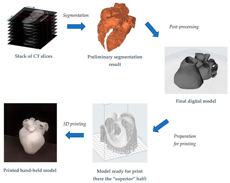
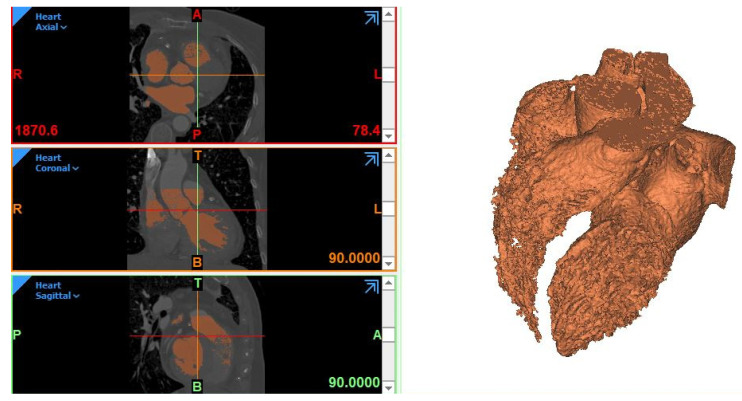
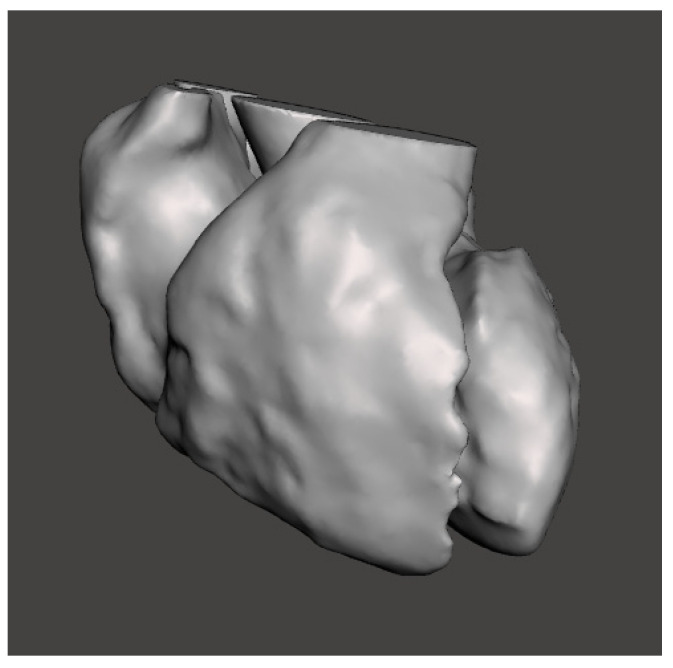
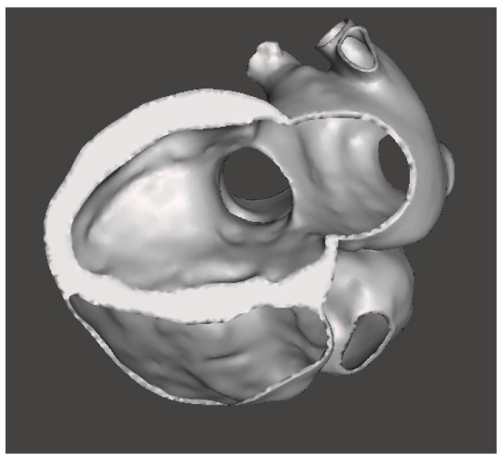
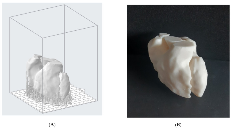
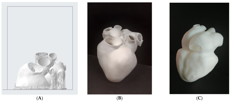
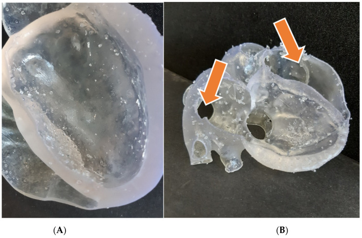
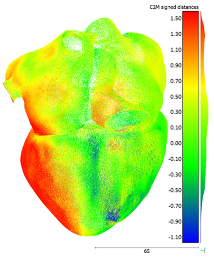
Medical images do not provide a natural visualization of 3D anatomical structures, while 3D digital models are able to solve this problem. Interesting applications based on these models can be found in the cardiovascular field. The generation of a good-quality anatomical model of the heart is one of the most complex tasks in this context. Its 3D representation has the potential to provide detailed spatial information concerning the heart's structure, also offering the opportunity for further investigations if combined with additive manufacturing. When investigated, the adaption of printed models turned out to be beneficial in complex surgical procedure planning, for training, education and medical communication. In this paper, we will illustrate the difficulties that may be encountered in the workflow from a stack of Computed Tomography (CT) to the hand-held printed heart model. An important goal will consist in the realization of a heart model that can take into account real wall thickness variability. Stereolithography printing technology will be exploited with a commercial rigid resin. A flexible material will be tested too, but results will not be so satisfactory. As a preliminary validation of this kind of approach, print accuracy will be evaluated by directly comparing 3D scanner acquisitions to the original Standard Tessellation Language (STL) files.
Keywords: 3D printing; heart model; patient-specific modeling; segmentation; stereolithography.
Conflict of interest statement
All authors declare no conflict of interest.
Figures
References
-
- Coppini G., Diciotti S., Valli G. Bioimmagini. Pàtron Editore; Bologna, Italy: 2012.
-
- Robinson R., Valindria V.V., Bai W., Oktay O., Kainz B., Suzuki H., Sanghvi M.M., Aung N., Paiva J.M., Zemrak F., et al. Automated quality control in image segmentation: Application to the UK Biobank cardiovascular magnetic resonance imaging study. J. Cardiovasc. Magn. Reson. 2019;21:1–14. doi: 10.1186/s12968-019-0523-x. - DOI - PMC - PubMed
-
- Bade R., Haase J., Preim B. SimVis. Citeseer; Princeton, NJ, USA: 2006. Comparison of Fundamental Mesh Smoothing Algorithms for Medical Surface Models; pp. 289–304.
LinkOut - more resources
Full Text Sources
Research Materials
