

Multifocality and Multicentrality in Breast Cancer: Comparison of the Efficiency of Mammography, Contrast-Enhanced Spectral Mammography, and Magnetic Resonance Imaging in a Group of Patients with Primarily Operable Breast Cancer
- PMID: 34677259
- PMCID: PMC8534697
- DOI: 10.3390/curroncol28050341
Multifocality and Multicentrality in Breast Cancer: Comparison of the Efficiency of Mammography, Contrast-Enhanced Spectral Mammography, and Magnetic Resonance Imaging in a Group of Patients with Primarily Operable Breast Cancer
Abstract
Background: The multifocality and multicentrality of breast cancer (MFMCC) are the significant aspects that determine a specialist's choice between applying breast-conserving therapy (BCT) or performing a mastectomy. This study aimed to assess the usefulness of mammography (MG), contrast-enhanced spectral mammography (CESM), and magnetic resonance imaging (MRI) in women diagnosed with breast cancer before qualifying for surgical intervention to visualize other (additional) cancer foci.
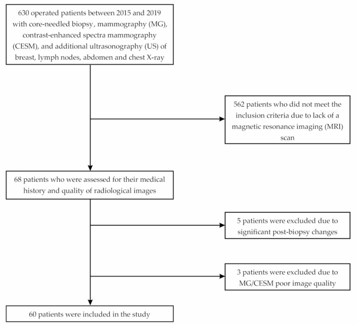
Methods: The study included 60 breast cancer cases out of 630 patients initially who underwent surgery due to breast cancer from January 2015 to April 2019. MG, CESM, and MRI were compared with each other in terms of the presence of MFMCC and assessed for compliance with the postoperative histopathological examination (HP).
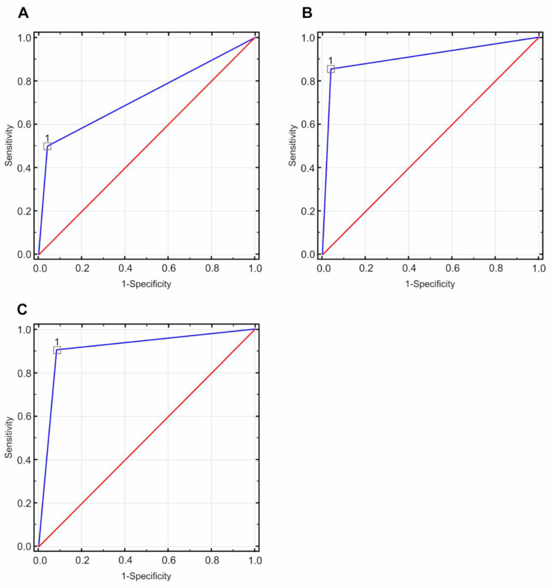
Results: Histopathological examination confirmed the presence of MFMCC in 33/60 (55%) patients. The sensitivity of MG in detecting MFMCC was 50%, and its specificity was 95.83%. For CESM, the sensitivity was 85.29%, and the specificity was 96.15%. For MRI, all the above-mentioned parameters were higher as follows: sensitivity-91.18%; specificity-92.31%.
Conclusions: In patients with MFMCC, both CESM and MRI are highly sensitive in the detection of additional cancer foci. Both CESM and MRI change the extent of surgical intervention in every fourth patient.
Keywords: breast cancer; comparative studies; contrast-enhanced spectral mammography; mammography; multifocal and multicentral breast cancers; pathology; surgery.
Conflict of interest statement
The authors declare no conflict of interest.
Figures
References
-
- Fahad Ullah M. Breast cancer: Current perspectives on the disease status. Advances in experimental medicine and biology. In: Ahmad A., editor. Breast Cancer Metastasis and Drug Resistance. Volume 1152. Springer International Publishing; Cham, Switzerland: 2019. pp. 51–64. - PubMed
-
- Fancellu A., Sanna V., Cottu P., Feo C.F., Scanu A.M., Farina G., Bulla A., Spanu A., Paliogiannis P., Porcu A. Mastectomy patterns, but not rates, are changing in the treatment of early breast cancer. Experience of a single european institution on 2315 consecutive patients. Breast. 2018;39:1–7. doi: 10.1016/j.breast.2018.02.003. - DOI - PubMed
-
- Nash R., Goodman M., Lin C.C., Freedman R.A., Dominici L.S., Ward K., Jemal A. State variation in the receipt of a contralateral prophylactic mastectomy among women who Received a diagnosis of invasive unilateral early stage breast cancer in the united states, 2004–2012. JAMA Surg. 2017;152:648. doi: 10.1001/jamasurg.2017.0115. - DOI - PMC - PubMed
-
- Veronesi U., Cascinelli N., Mariani L., Greco M., Saccozzi R., Luini A., Aguilar M., Marubini E. Twenty-year follow-up of a randomized study comparing breast-conserving surgery with radical mastectomy for early breast cancer. N. Engl. J. Med. 2002;347:1227–1232. doi: 10.1056/NEJMoa020989. - DOI - PubMed
-
- Hartmann-Johnsen O.J., Kåresen R., Schlichting E., Nygård J.F. Better survival after breast-conserving therapy compared to mastectomy when axillary node status is positive in early stage breast cancer: A registry-based follow-up study of 6387 Norwegian women participating in screening, primarily operated between 1998 and 2009. World J. Surg.Onc. 2017;15:118. doi: 10.1186/s12957-017-1184-6. - DOI - PMC - PubMed
MeSH terms
Substances
LinkOut - more resources
Full Text Sources
Medical
Research Materials
Miscellaneous
