Review
. 2021 Oct 12;11(10):385.
doi: 10.3390/bios11100385.

Applications of Microfluidics in Liquid Crystal-Based Biosensors

Affiliations
Review

Applications of Microfluidics in Liquid Crystal-Based Biosensors

Jinan Deng et al. Biosensors (Basel). .

Abstract

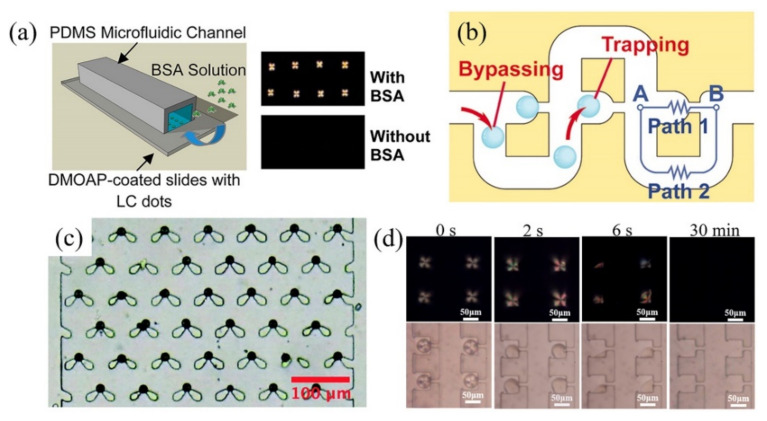
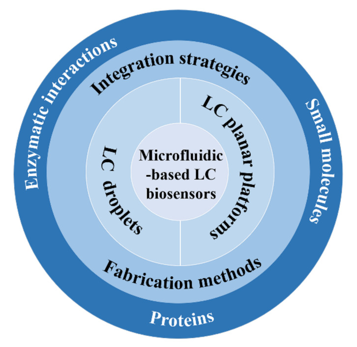
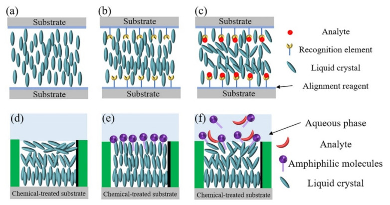
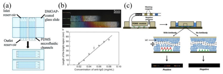
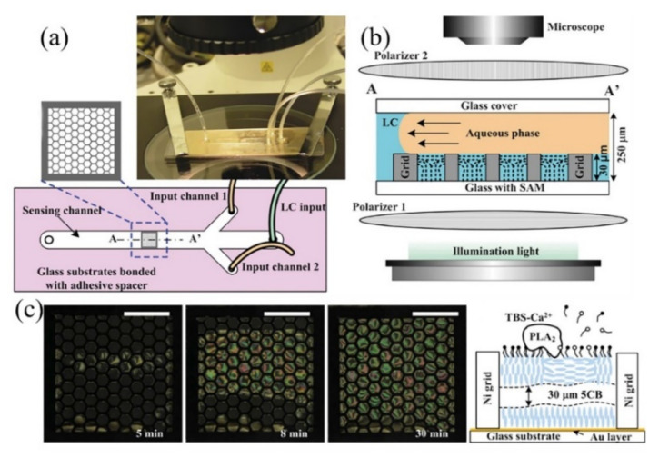
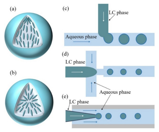
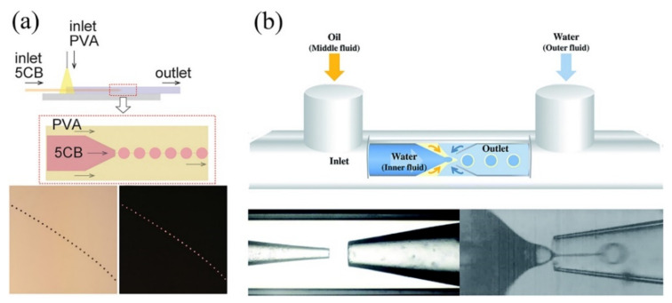
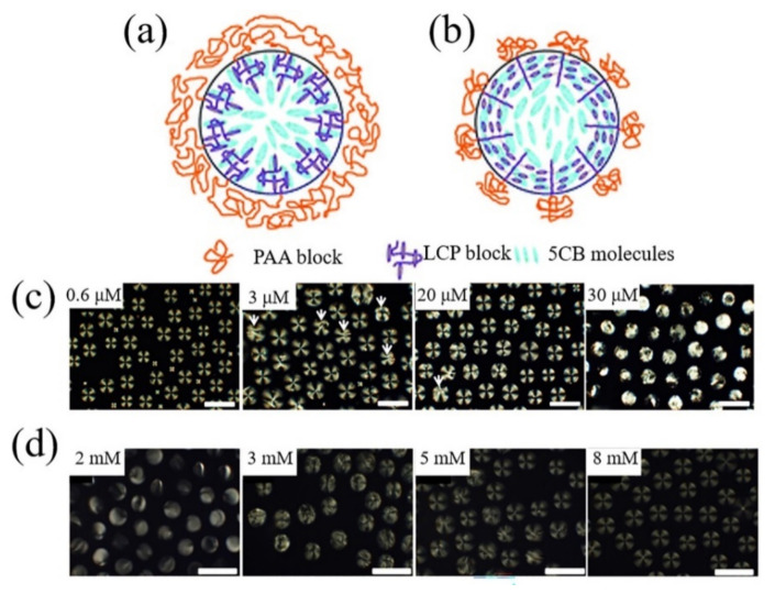
Liquid crystals (LCs) with stimuli-responsive configuration transition and optical anisotropic properties have attracted enormous interest in the development of simple and label-free biosensors. The combination of microfluidics and the LCs offers great advantages over traditional LC-based biosensors including small sample consumption, fast analysis and low cost. Moreover, microfluidic techniques provide a promising tool to fabricate uniform and reproducible LC-based sensing platforms. In this review, we emphasize the recent development of microfluidics in the fabrication and integration of LC-based biosensors, including LC planar sensing platforms and LC droplets. Fabrication and integration of LC-based planar platforms with microfluidics for biosensing applications are first introduced. The generation and entrapment of monodisperse LC droplets with different microfluidic structures, as well as their applications in the detection of chemical and biological species, are then summarized. Finally, the challenges and future perspectives of the development of LC-based microfluidic biosensors are proposed. This review will promote the understanding of microfluidic techniques in LC-based biosensors and facilitate the development of LC-based microfluidic biosensing devices with high performance.

Keywords: biosensors; liquid crystal droplets; liquid crystal planar interface; liquid crystals; microfluidics.


Conflict of interest statement

The authors declare no conflict of interest.

Figures

Figure 7
Figure 7
(a) LC droplet-based static microarray for the detection of BSA. Reprinted with permission from ref. [54]. Copyright 2013 American Chemical Society. (b) The schematic diagram of the trap mechanism using by-pass microfluidic channel. Reprinted with permission from ref. [100]. Copyright 2007 National Academy of Science of the USA. (c) Optical microscopy image of LC droplets trapped in a swallowtail shaped microstructure. Reprinted with permission from ref. [57]. Copyright 2019 Royal Society of Chemistry. (d) Polarizing optical microscopy and corresponding bright-field microscopy images of LC droplet-based microarray after the introduction of 20 μM cholic acid at a constant flow rate of 3 μL/h. Reprinted with permission from Reference [55]. Copyright 2020 Elsevier.
Scheme 1
Scheme 1
Schematic illustration of the contents in this review.
Figure 1
Figure 1
Schematic illustrations of the alignment of LCs on (a) alignment reagent-treated substrate, (b) recognition element-decorated alignment reagent-treated substrate and (c) recognition element-decorated alignment reagent-treated substrate after the addition of analyte. Schematic illustrations of LC-aqueous interface (d) without and (e) with the assembled amphiphilic molecules. (f) The amphiphilic molecules-decorated LC-aqueous interface after the addition of analyte.
Figure 2
Figure 2
(a) Schematic illustrations of the preparation of LC-solid interface with microfluidic techniques for multiplex analysis. Reprinted with permission from ref. [69]. Copyright 2011 American Chemical Society. (b) Polarized optical microscopy images of IgG-decorated LC-solid interface-based microfluidic platform after the addition of anti-IgG of (from top) 0.02 mg/mL, 0.05 mg/mL, and 0.08 mg/mL, respectively, and the length of the bright region as a function of the anti-IgG concentration. Reprinted with permission from ref. [68]. Copyright 2009 John Wiley and Sons. (c) Schematic illustrations of the working principle of one-step LC-based microfluidic immunoassay. Reprinted with permission from ref. [67]. Copyright 2014 Elsevier.
Figure 3
Figure 3
(a) Schematic illustration and photograph of microfluidic device for the fabrication of LC-aqueous interface. (b) Schematic illustration of the formation of LC-aqueous interface within the microchannel. (c) The gradual optical appearance transitions of L-DLPC-decorated LC-aqueous interface within the microchannel after the addition of phospholipase A2 in the presence of Ca2+ and the related LC ordering transition mechanism. Reprinted with permission from ref. [51]. Copyright 2012 Royal Society of Chemistry.
Figure 4
Figure 4
Schematic illustrations of (a) bipolar and (b) radial configurations of LC droplets. Schematic illustrations of generation of monodisperse LC droplets with (c) T-junction, (d) flow-focusing and (e) co-flow microstructures.
Figure 5
Figure 5
(a) LC necklace structure prepared by co-flow microstructure. Reprinted with permission from ref. [93]. Copyright 2020 American Chemical Society. (b) LC double emulsion droplets produced by the combination of co-flow and flow-focusing geometries. Reprinted with permission from ref. [89]. Copyright 2016 Royal Society of Chemistry.
Figure 6
Figure 6
Configurations of PAA-b-LCP-decorated LC droplet at (a) higher pH and (b) lower pH. Reprinted with permission from ref. [52]. Copyright 2011 Royal Society of Chemistry. (c) The polarized optical microscopy images of GOx-coated LC droplets with different glucose concentrations. Reprinted with permission from ref. [27]. Copyright 2013 American Chemical Society. (d) The polarized optical microscopy images of urease-coated LC droplets with different urea concentrations. Reprinted with permission from ref. [95]. Copyright 2014 Elsevier. The scale bar is 100 μm.

Similar articles

Cited by

References

    1. He Z., Gou F., Chen R., Yin K., Zhan T., Wu S.T. Liquid crystal beam steering devices: Principles, recent advances, and future developments. Crystals. 2019;9:292. doi: 10.3390/cryst9060292. - DOI
    1. Bisoyi H.K., Li Q. Light-driven liquid crystalline materials: From photo-induced phase transitions and property modulations to applications. Chem. Rev. 2016;116:15089–15166. doi: 10.1021/acs.chemrev.6b00415. - DOI - PubMed
    1. Bremer M., Kirsch P., Klasen-Memmer M., Tarumi K. The TV in your pocket: Development of liquid-crystal materials for the new millennium. Angew. Chem. Int. Ed. 2013;52:8880–8896. doi: 10.1002/anie.201300903. - DOI - PubMed
    1. Woltman S.J., Jay G.D., Crawford G.P. Liquid-crystal materials find a new order in biomedical applications. Nat. Mater. 2007;6:929–938. doi: 10.1038/nmat2010. - DOI - PubMed
    1. Prakash J., Parveen A., Mishra Y.K., Kaushik A. Nanotechnology-assisted liquid crystals-based biosensors: Towards fundamental to advanced applications. Biosens. Bioelectron. 2020;168:112562. doi: 10.1016/j.bios.2020.112562. - DOI - PubMed

LinkOut - more resources