

Posterior Vitreous Detachment in Normal Healthy Subjects Younger Than Age Twenty
- PMID: 34677570
- PMCID: PMC8543394
- DOI: 10.1167/iovs.62.13.19
Posterior Vitreous Detachment in Normal Healthy Subjects Younger Than Age Twenty
Abstract
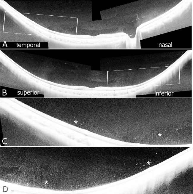
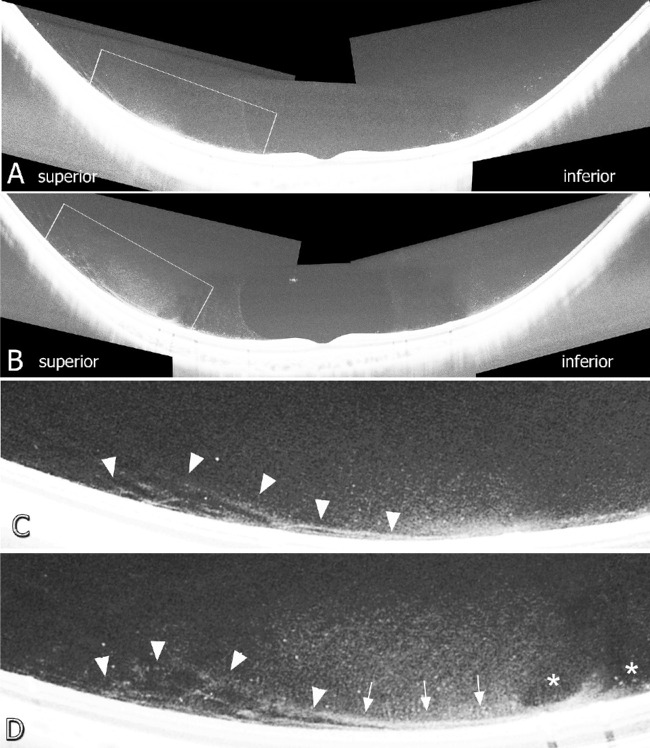
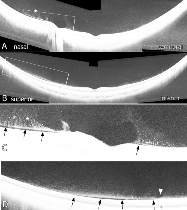
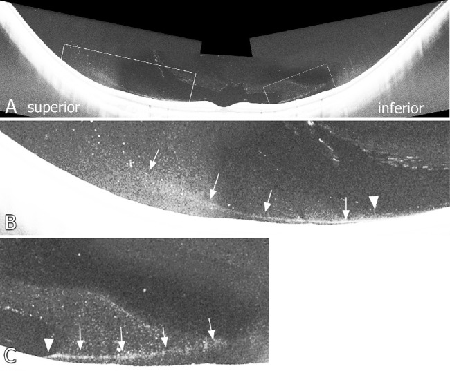
Purpose: To describe the initiation of posterior vitreous detachment (PVD) in the eyes of normal individuals, under 20 years of age, using wide-angle optical coherence tomography (OCT).
Methods: This is an observational cross-sectional study. Montaged images of horizontal and vertical OCT scans were obtained in 63 healthy eyes of 35 consecutive subjects ranging in age from 4 to 17 years.
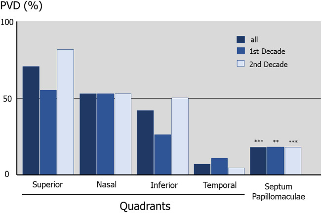
Results: Forty-five eyes (71.4%) had obvious PVD, defined as a contiguous line of posterior cortical vitreous separated from the surface of the retina. Eighteen eyes (28.6%) had no PVD. The mean age of the individuals without PVD was significantly younger than those with PVD (P = 0.008). The spatial distribution of PVD initiation was highest in the superior quadrants, with the nasal, inferior, septum papillomaculae, and temporal quadrants following in descending order of frequency (P < 0.001). PVD was observed to begin anterior to the premacular liquefied lacuna, where the vitreous gel directly adheres to the vitreoretinal interface. In the majority of subjects (80.6%), PVD was initiated anterior to the vascular arcades.
Conclusions: PVD can be observed by OCT to begin in the first and second decade of life. It begins in the mid-peripheral vitreous, most frequently in the superior quadrants anterior to the vascular arcades. In this study, all PVDs originated outside of the macular liquefied lacunae, where the vitreous gel adheres directly to the retina.
Conflict of interest statement
Disclosure:
Figures
References
-
- Tozer K, Johnson MW, Sebag J.. Vitreous aging and posterior vitreous detachment. In: Sebag J, ed. The Vitreous in Health and Disease. New York: Springer; 2014: 131–150.
-
- Johnson MW. Posterior vitreous detachment: evolution and role in macular disease. Retina. 2012; 32(suppl): S174–S178. - PubMed
-
- Uchino E, Uemura A, Ohba N.. Initial stages of posterior vitreous detachment in healthy eyes of older persons evaluated by optical coherence tomography. Arch Ophthalmol. 2001; 119(10): 1475–1479. - PubMed
-
- Johnson MW. Posterior vitreous detachment: evolution and complications of its early stages. Am J Ophthalmol. 2010; 149(3): 371–382. - PubMed
Publication types
MeSH terms
LinkOut - more resources
Full Text Sources
