Review
. 2022 Mar 9;122(5):5233-5276.
doi: 10.1021/acs.chemrev.1c00365. Epub 2021 Oct 22.

Dissecting Biological and Synthetic Soft-Hard Interfaces for Tissue-Like Systems

Affiliations
Review

Dissecting Biological and Synthetic Soft-Hard Interfaces for Tissue-Like Systems

Yin Fang et al. Chem Rev. .

Abstract

Soft and hard materials at interfaces exhibit mismatched behaviors, such as mismatched chemical or biochemical reactivity, mechanical response, and environmental adaptability. Leveraging or mitigating these differences can yield interfacial processes difficult to achieve, or inapplicable, in pure soft or pure hard phases. Exploration of interfacial mismatches and their associated (bio)chemical, mechanical, or other physical processes may yield numerous opportunities in both fundamental studies and applications, in a manner similar to that of semiconductor heterojunctions and their contribution to solid-state physics and the semiconductor industry over the past few decades. In this review, we explore the fundamental chemical roles and principles involved in designing these interfaces, such as the (bio)chemical evolution of adaptive or buffer zones. We discuss the spectroscopic, microscopic, (bio)chemical, and computational tools required to uncover the chemical processes in these confined or hidden soft-hard interfaces. We propose a soft-hard interaction framework and use it to discuss soft-hard interfacial processes in multiple systems and across several spatiotemporal scales, focusing on tissue-like materials and devices. We end this review by proposing several new scientific and engineering approaches to leveraging the soft-hard interfacial processes involved in biointerfacing composites and exploring new applications for these composites.


Figures

Figure 1.
Figure 1.
Soft–hard interfaces cover a wide range of length and time scales. (a) The interfaces are diverse and cover those in molecular crystals, nanocrystal superlattices, nanowire nucleation and growth, biomineralization in both healthy and pathological conditions, active battery interfaces, soil chemistry and geobiology, and bioelectronics and implants. The soft and hard components are represented by colors within the range of the respective color bars (upper right). While the overall length scale ranges from subnanometer to above centimeter, the critical interfacial processes all occur at the molecular and nanometer scales where the interfacial mismatches are minimal. (b) Time scale is broad, and includes mechanical, electrical, and chemical events (shown here), such as bond vibration and enzyme turnover.
Figure 2.
Figure 2.
Several figures-of-merit for material mechanics are relevant to this review. (a) Young’s moduli of naturally occurring and synthetic materials. (b) Stress–strain curves and a few relevant definitions.
Figure 3.
Figure 3.
Soft–hard interfacial processes have essential roles. (a) The interfacial process can yield new morphologies difficult to obtain with soft or hard components alone. It can stabilize the components against fragmentation, degradation, or disintegration. It also enables signal transductions between the soft and hard components. (b) 3D reconstructed cryo-TEM image of a dodecahedral silica cage, showing the intricate 3D architecture of the interface and the nanoscale feature size. The cryo-TEM image is modified with permission from ref . Copyright 2018 Springer Nature. (c) The high curvature from nanopillar structures recruits multiple curvature-sensing proteins, with protein names shown on the right. The image is modified with permission from ref . Copyright 2017 Springer Nature.
Figure 4.
Figure 4.
Processes at soft–hard interfaces are unique. (a) Although different energies display different dependence on object size or dimensions, they converge at the molecular and nanoscopic scale. At this length scale, efficient signal transduction and minimal mismatches are expected at soft–hard interfaces. At this size regime, the boundaries between soft and hard materials, and the boundaries between living and nonliving systems, become blurred. This energy convergence can explain the critical roles of nanoscale interfacial processes at many soft–hard interfaces. Reproduced with permission from ref . Copyright 2019 Nature Springer. (b) Dendritic binders and nanoclays self-assemble to form stable hydrogel. The feature sizes of the building blocks are at the molecular and nanoscopic levels, where multiple energy terms show similar amplitude such that the soft–hard interfaces can establish in a seamless manner. (c) To mitigate interfacial mismatches, additional buffer mechanisms are available. These mechanisms include the use of connecting linkers and deposits (additive strategy), the formation of a modulus gradient over the distance between the soft and hard phases (spatial strategy), and gradual and less perturbative condensation of the hard component at the interface (temporal strategy). (d) The soft–hard interfaces can produce the strain hotspots, which may be leveraged for special chemical processes or applications. For example, ring sliding of polyrotaxane close to the silicon/binder interfaces can be triggered during the operation of silicon-based battery anode.
Figure 5.
Figure 5.
Soft–hard interfaces present several pathways of exploring new interfacial processes, by either mitigating or leveraging the mismatches at the interfaces. Shown here only highlights the chemical processes.
Figure 6.
Figure 6.
Multiple characterization approaches have been developed to study the dynamic interfacial processes at the soft–hard interfaces. (a) In situ studies of short processes can be achieved with ultrafast atomic force microscopy (AFM), environmental transmission electron microscopy (TEM), small angle X-ray scattering (SAXS) and confocal laser scanning microscopy (CLSM). (b) While in situ experiments can capture the dynamics of short processes directly, ex situ studies can reveal the dynamics indirectly by recording static information at multiple time points (blue dots alone the dashed line arrow), with correlative microscopies and spectroscopies, and with input from theory and simulation. SEM: scanning electron microscope; TEM: transmission electron microscope; AFM: atomic force microscope; CLSM: confocal laser scanning microscopy; XRD: X-ray diffraction; SAXS: Small-angle X-ray scattering; SAED: selected area electron diffraction; STEM: scanning transmission electron microscopy; MD: molecular dynamics simulation; PCR: polymerase chain reaction; SDS-PAGE: sodium dodecyl sulfate polyacrylamide gel electrophoresis; IHC: immunohistochemistry; XPS: X-ray photoelectron spectroscopy; NMR: Nuclear magnetic resonance; FTIR; Fourier transform infrared spectroscopy; EDS: energy-dispersive X-ray spectroscopy; EELS: electron energy loss spectroscopy; APT: atom probe tomography; SIMS: secondary-ion mass spectrometry.
Figure 7.
Figure 7.
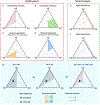
We propose a new “Interaction Framework” at the soft–hard interfaces. The interactions can be (bio)chemical, mechanical, electrical, etc. (a–d) represent the spatial aspect, while panels e and f represent the temporal aspect of the interaction framework. Depending on the specific target/domain of a system, different interaction frameworks can coexist in a single system. (g) Similar to the interpretation of a conventional phase diagram, the relative distance from a point (blue dot) to the three corners of the framework triangle suggests the relative dominance of the roles of SS, SH, and HH interactions; the shorter the distance to one corner, the more dominant the interaction that is represented by the corner. Each color domains in a–d are the overlapped area from two half-triangles (in gray) as two inequalities need to be satisfied simultaneously. The approach to derive the domain for the case of “deterministic assembly” is given in panel g.
Figure 8.
Figure 8.
Interaction framework–cooperative integration can be verified with existing examples. (a) The cooperative integration diagram. (b) Synthesis of mesostructured inorganic materials often involves self-assembly of inorganic precursors (positively charged I+ or negatively charged I) with cationic surfactants (S+) or nonionic block copolymers (N0). The soft–hard (SH) interactions can involve protonation (H+) and anion coupling (X), and occur in either acidic or basic conditions. In block copolymer-based synthesis, e.g., with pluronic F127, the hydrophilic segments such as PEO are inserted inside the inorganic walls, producing a graded regime. The addition of cosurfactant such as sodium bis(2-ethylhexyl) sulfosuccinate (AOT) can tune the interface chemistry and the curvature. (c) Electrochemical intercalation of black phosphorus (BP) by CTAB molecules produces a soft–hard superlattice structure. Electrochemical reactions are shown in the lower left panel. As the superstructure cannot be formed by either the black phosphorus or the CTAB molecules alone, the cooperativity at the soft–hard interfaces is the key to the final composite structure.
Figure 9.
Figure 9.
Interaction framework–directed organization from the soft phase can be verified with existing examples. (a) The interaction framework for the soft phase-directed organization. (b) In a naturally occurring example, the production of chitin-binding proteins (DgCBPs) and histidine-rich proteins (DgHBPs) based coacervates, the dynamic interaction of these proteins with the chitin-based soft template, and hydration/dehydration create the modulus gradient. Panels c–e are synthetic examples. (c) Schematic of the strategy enabling programmed deposition of electrostatic charges. Patterns of positive, negative, and neutral patterns allow for selective material deposition and preserve the prescribed charge in 3D materials. Reproduced with permission from ref . Copyright 2020 Springer Nature. (d) The optically patternable hydrogels triggered volumetric metal deposition and intensification at defined 3D locations. Upon volume shrinkage, mineralized networks such as a silver nanostructure and optical metamaterials can be generated. Reproduced with permission from ref. Copyright 2018 AAAS. (e) Liquid alloy droplets or liquid metal droplets can template the growth of 2D inorganic sheets over their surfaces. The droplets also serve as the chemical reactors where at least one chemical element is extracted from the droplets for the sheet production. ΔG(S) is the free energy change for the solid skin. NAu and NSi are the area density of Au and Si atoms in the skin layer. ΔΓ(S) is the change in free energy upon taking NAu + NSi atoms from the reference solids. μAu(l) and μSi(l) are the chemical potentials of the atoms in the liquid.
Figure 10.
Figure 10.
Interaction framework–directed organization from the hard phase can be verified with existing examples. (a) The interaction framework for the hard phase-directed organization. (b) De novo designed proteins form ordered structures over the mica surface. The upper schematic shows the model of DHR-mica interface, with repeats 1–3 (glutamate, Glu, side chains), repeats 4–6 (α-helical secondary structures), repeats 7–9 (full DHR backbone), repeats 10–12 (backbone and amino acid side chains), repeats 13–15 (all atoms), and repeats 16–18 (external protein surface). The lower AFM image shows one honeycomb lattice. The inset area is 200 nm × 200 nm. This panel is adapted with permission from ref . Copyright 2019 Springer Nature.
Figure 11.
Figure 11.
Interaction framework–signal transduction can be verified with existing examples. (a) The interaction framework for the hard phase-initiated signal transduction. (b) The formation of kidney crystal granulomas is an example of cooperative and multiple interfacial processes in a soft–hard hybrid generation. (c) A photoelectrochemical process at a coaxial silicon nanowire/neuron interface can elicit action potential propagation in cells. Adapted with permission from ref . Copyright 2018 Springer Nature.
Figure 12.
Figure 12.
Several strategies have been developed to produce soft tissue-like materials. (a) Self-growing muscle-like hydrogels. Reproduced with permission from ref . Copyright 2019 AAAS. (b) Granules-enabled tissue-like materials from granule/hydrogel composite. Reproduced with permission from ref . Copyright 2020 ELSEVIER. (c) A multilength-scale hierarchical hydrogel architecture that mimicks tendon was made by directional freezing and salting out methods. Reproduced with permission from ref . Copyright 2021 Springer Nature.
Figure 13.
Figure 13.
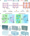
Interaction between soft substrates and hard electronics produces stretchable and three-dimensional devices. (a) Interaction between the stretchable substrate and silicon material allows the formation of highly uniform ribbons. Reproduced with permission from ref . Copyright 2006 AAAS. (b) Cooperative integration, SH > SS, SH > HH. The wavy structure was formed through a biphasic cooperative process. This structure cannot form with PDMS or Si nanoribbon alone. (c) Arrays of three-dimensional multifunctional mesoscale frameworks for holding and recording electrical activity in spheroid organoids. Reproduced with permission from ref . Copyright 2021 AAAS. (d) Directed organization, SS > SH > HH. The local bonding sites and the elastomer substrate enable 2D-3D transformation. (e) Stress between the hard (metal) and soft (SU-8) materials allows for transistor positioning in a recording device. Reproduced with permission from ref . Copyright 2012 Springer Nature. (f) Directed organization, HH > SH > SS. The residual stress from multiple material layers enables the 2D–3D transformations.
Figure 14.
Figure 14.
Atomically thin-material interlayers enable strain-resilient electrodes. (a) Schematic of a fractured metal film on single-layer graphene forming the conductive and flexible electrode. (b) Conceptual plot comparing resistance in bare metal and metal-interlayer material in the function of applied strain. The metal-interlayer material possesses a large, stable “resistance locking” region. (c) Optical image of a flexible transistor array under 0% (left) and 100% (right) deformation. The varying concentration of cross-linkers in conductive polymers allows for the fabrication of the device with the prescribed stiffness allowing redistribution of strain to the nonactive areas. (d) Representative structure of a flexible transistor device. (SC, semiconductor material) Panels a and b were reproduced with permission from ref . Copyright 2021 Springer Nature. Panels c and d reproduced with permission from ref . Copyright 2021 Springer Nature.
Figure 15.
Figure 15.
Flexible ultrasonic device has high mechanical resilience and can conform to the skin enabling remote sensing of blood pressure. (a) Schematic of the ultrasonic device with the array of piezoelectric rods embedded in an epoxy matrix. (b) Optical image of the device under strain showing its high conformability. (c) The device was made by the deterministic assembly, where the properties of the soft and hard phases are well-defined. Reproduced with permission from ref . Copyright 2018 Springer Nature.
Figure 16.
Figure 16.
Self-healing elastomers enable regeneration in electronic systems. (a) Schematic of hydrogen bonding combinations in the self-healable elastomer. Reproduced with permission from ref . Copyright 2018 Wiley. (b) Schematic of the autonomous resistance recovery in the carbon nanotube (CNT) network embedded in the self-healing polymer. (1) In the original state, the material possesses high conductivity due to interweaving CNTs in the network. (2) Mechanical damage (e.g., cut) increases the material’s resistance. (3) Mechanical stability and conductivity are regained after contact between cut parts is established. (4) Due to the dynamic nature of the polymer, the CNT network recovers, and the cut part is indistinguishable from the pristine material. Adapted with permission from ref . Copyright 2018 Springer Nature. (c) Directed organization following SS > SH > HH can be used to describe the self-healing behavior of the 1D nanostructure-incorporated PDMS elastomer.
Figure 17.
Figure 17.
Electronic inks enable the printing of electronic circuits directly on the skin and achieve unprecedented conformity. (a) A demonstration of an electronic ink deposition using polyimide stencil. (b) The material shows direct organization on the surface of the skin, following the directed organization pathway. (c) Drawn on skin electronics have been used to promote wound healing through electrical stimulation. The images show accelerated healing in the treated region (top part of the wound) compared to the untreated region (bottom part of the wound). Reproduced with permission from ref . Copyright 2020 Springer Nature.
Figure 18.
Figure 18.
Bioinspired architecture allows to produce minimally invasive and biocompatible neuron-like electrodes. (a) Schematics of neuron-like electronics (NeuE) and neurons showing their structural similarity at the subcellular level and the network level (inset). NeuE is fabricated using photolithography. Submicrometer-thick and a few micrometer-wide neurite-like SU-8/gold/SU-8 interconnects are connected to platinum electrodes. (b) Two-photon fluorescence image of the interface between neurons (green) and NeuE (red) at 6 weeks after injection into the mouse brain. The white dashed circle indicates an electrode. (c) Bar chart showing the number of neurons recorded by each electrode as a function of postimplantation time. Bar colors are coded according to the brain regions spanning from the cortex (CTX) to hippocampal CA1 and CA3. (d) While device fabrication follows deterministic assembling, the signal transduction for neural recording starts from a different soft system, i.e., neural tissues. Panels a–c are reproduced with permission from ref . Copyright 2020 Springer Nature.
Figure 19.
Figure 19.
Organogenesis can guide an organization of 3D mesh electronics. (a) Microscope images showing different types of deformations in the material throughout organogenesis. (b) Directed organization, following SS > SH > HH. Organoid development provided the driving force for the 2D to 3D device transformation. (c) Seamless integration enabled continuous chronic recording and analysis of spike dynamics revealed a change from an initially slow waveform to fast depolarization over the course of organogenesis. Reproduced with permission from ref . Copyright 2019 ACS.
Figure 20.
Figure 20.
Silicon nanowires can be spontaneously internalized by different types of cells, enabling optical modulation of cells. (a) Confocal microscope image of nanowires (blue, scattering channel) internalized by HUVEC cells. The confocal sections confirm nanostructure internalization. (b) The plot of area overlap between the nanowires and cell bodies and confluence over time. Overlap fraction larger than confluence suggests that nanowires are actively taken up by the cells. (c) soft–hard interaction framework follows SH > SS and SH > HH. (d) DIC microscopy image of nanowires internalized by glial cells. (black arrow–nanowire, red arrow–glial cell, blue arrow–neuron) Confocal microscope fluorescence time series shows calcium wave propagation after stimulation of an internalized nanowire using a laser. (e) Transients of calcium dynamics in the cellular assembly after stimulation. (f) Schematic of myofibroblast-cardiomyocytes assembly, which can be used to study intercellular signaling and perform pacing of muscle cells. Panels a and b were reproduced with permission from ref . Copyright 2016 AAAS. Panels d and e reproduced with permission from ref . Copyright 2018 Springer Nature. Panel f reproduced with permission from ref . Copyright 2019 PNAS.
Figure 21.
Figure 21.
Genetic encoding of the catalytic enzyme allows the synthesis of conductive polymers directly onto the cell membranes. (a) Genetic targeting allows modifying only a specific subset of neurons with the conductive polymer. Depending on the polymer conductivity, these materials can enhance or inhibit neuronal activity by modifying their membranes’ capacitance. Reproduced with permission from ref . Copyright 2020 AAAS. (b) Directed organization following SS > SH> HH. Genetically modified neurons can guide the growth of the conducting polymers. In this case, the modulus of the conducting polymers are orders of magnitude larger than that of cell membranes. This process can be homologous to biomineralization, although no inorganic materials are formed.
Figure 22.
Figure 22.
Phototactic gels allow the creation of oscillators that can be used for light-driven motion. (a) Schematic of the laser-driven oscillating pillar made of the phototactic gel. (b) soft–hard interaction framework. The signal transduction follows HH > SH > SS. (c) Mechanism of oscillating motion. Self-shadowing of the pillar’s inner surface creates out-of-equilibrium condition and negative loop driving the reciprocal motion. (d) Tip displacement and local temperature in the hinge during motion. The 90° shift between the curves is in agreement with the proposed motion mechanism. Reproduced with permission from ref . Copyright 2019 AAAS.
Figure 23.
Figure 23.
Future exploration at soft–hard interfaces can yield numerous advances. (a) Bone formation presents several biomimetic chemical approaches. The chemical gradient in a tendon may suggest new ways of building load-bearing interfaces between implants and tissues. Transcortical vessels, the newly discovered trans-cortical capillaries, and the spongy structures indicate that building micro- or nanoscale networks within the hard-phase may improve mass transport to soft–hard interfaces. The biogenic synthesis and regulation of hydroxyapatites over the collagen matrix may address the need for greener and homeostatic chemical processes. (b) Property mismatches at soft–hard interfaces can be leveraged for unique chemical processes. For example, the “host–guest chemistry” from soft materials, when coupled with the deterministic electronic processing from the hard counterpart, may yield new chemical or biochemical sensor devices. Additionally, mismatches at interfaces can produce gradients and fields, which could trigger chemical or biochemical reactions in a highly efficient manner.
Figure 24.
Figure 24.
New strategies for tissue engineering would enable future biointegrated electronics and robotics. (a) Hydrogel-directed axonal growth was used to generate living electrodes. Optogenetically active neurons were grown in a cylinder. After implantation, rapid axonal overgrowth allowed integration of the probe with the tissue. Partial overgrowth of host axons with the probe allows for the recording of neuronal activity. (b) Schematic of two probes of various lengths which can be implanted to distinct depth allowing stimulation of different parts of the brain using LED arrays. (c) High throughput mechano-pharmacological screen was used to produce mesenchymal stem-cell-derived endothelial cells, which promote vascularization and perfusion in the tissue after transplantation. Both chemical and mechanical stimuli were applied to generate appropriate cell phenotype. Panels a and b were reproduced with permission from ref . Copyright 2021 AAAS. Panel c reproduced with permission from ref . Copyright 2021 Springer Nature.

Similar articles

Cited by

References

    1. Kato T; Yoshio M; Ichikawa T; Soberats B; Ohno H; Funahashi M Transport of ions and electrons in nanostructured liquid crystals. Nat. Rev. Mater 2017, 2, 1–20.
    1. Yang CH; Suo ZG Hydrogel ionotronics. Nat. Rev. Mater 2018, 3, 125–142.
    1. Wegst UGK; Bai H; Saiz E; Tomsia AP; Ritchie RO Bioinspired structural materials. Nat. Mater 2015, 14, 23–36. - PubMed
    1. Whittell GR; Hager MD; Schubert US; Manners I Functional soft materials from metallopolymers and metallosupramolecular polymers. Nat. Mater 2011, 10, 176–188. - PubMed
    1. Weiss RG The past, present, and future of molecular gels. what is the status of the field, and where is it going? J. Am. Chem. Soc 2014, 136, 7519–7530. - PubMed

Publication types

MeSH terms