. 2021 Oct 22;6(64):eabj3859.
doi: 10.1126/sciimmunol.abj3859. Epub 2021 Oct 22.

NLRP3 inflammasome activation triggers gasdermin D-independent inflammation

Affiliations

NLRP3 inflammasome activation triggers gasdermin D-independent inflammation

Chun Wang et al. Sci Immunol. .

Abstract

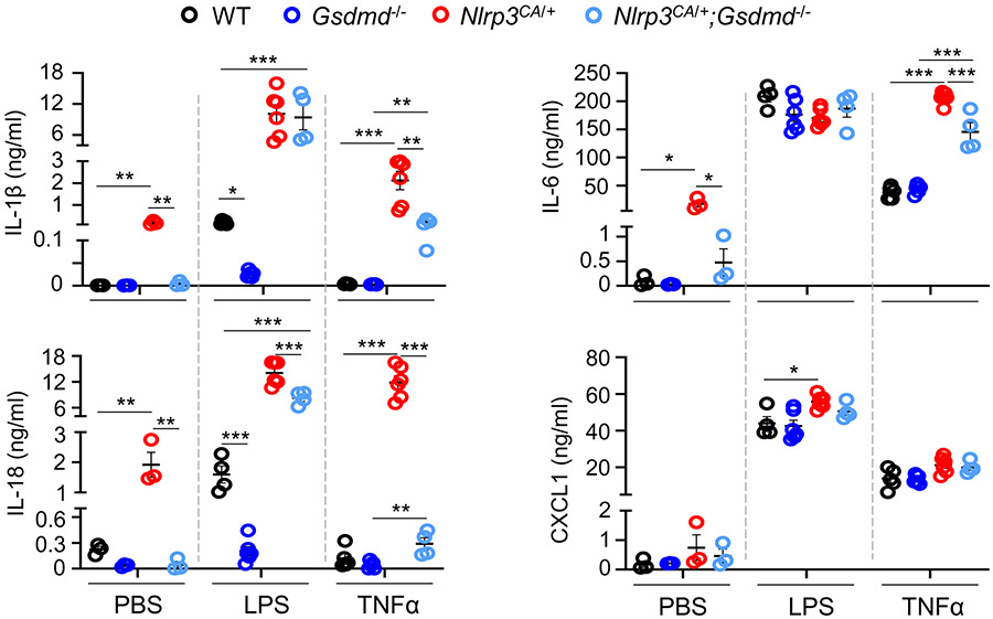
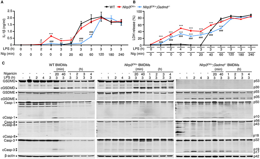
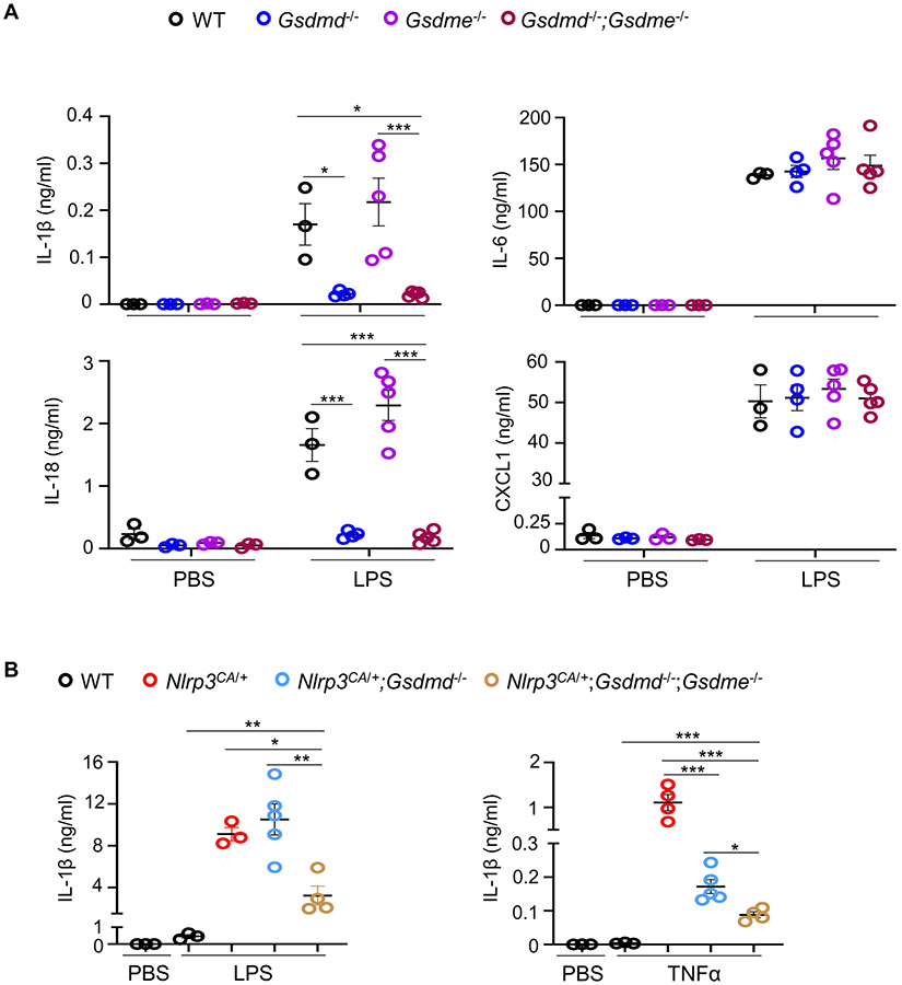
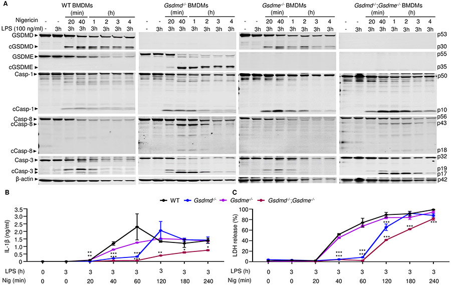
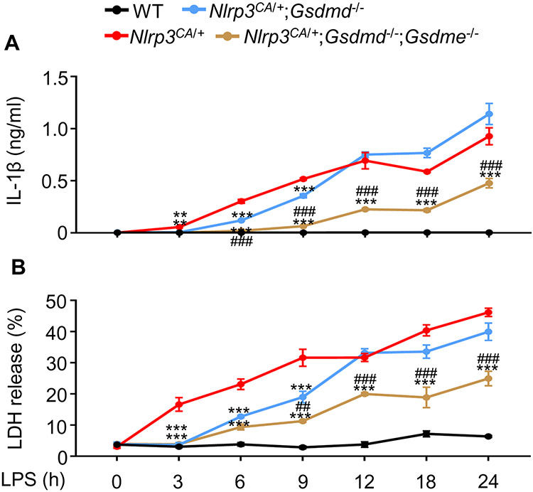
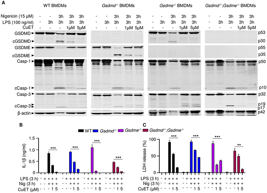
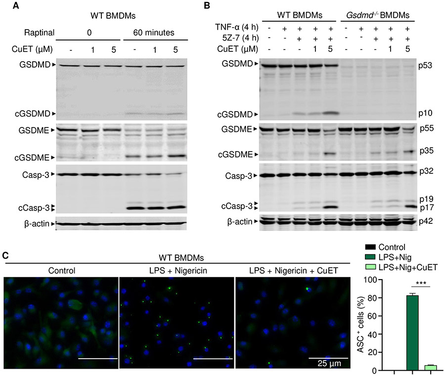
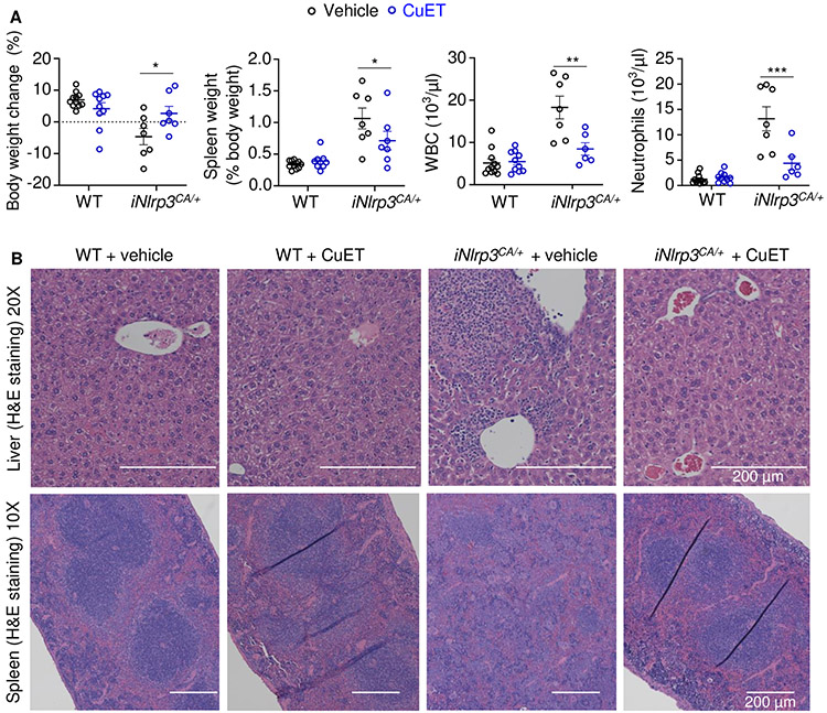
NOD-like receptor (NLR), family pyrin domain containing 3 (NLRP3) assembles a protein complex known as the NLRP3 inflammasome upon sensing certain pathogen products or sterile danger signals. Gain-of-function mutations such as the D301N substitution in NLRP3, which cause its constitutive activation (NLRP3CA) also results in inflammasome assembly. This inflammasome processes pro–interleukin-1 β (pro–IL-1β) and pro–IL-18 into bioactive IL-1β and IL-18, respectively, and cleaves gasdermin D (GSDMD). GSDMD amino-terminal fragments form plasma membrane pores that facilitate the secretion of IL-1β and IL-18 and lead to the inflammatory cell death pyroptosis. Accordingly, GSDMD inactivation results in negligible spontaneous inflammation in various experimental models such as in Nlrp3CA/+ mice lacking GSDMD (Nlrp3CA/+;Gsdmd−/− mice). Here, we found that Nlrp3CA/+;Gsdmd−/− mice, when challenged with LPS or TNF-α, still secreted IL-1β and IL-18, indicating inflammasome activation independent of GSDMD. Accordingly, Gsdmd−/− macrophages failed to secrete IL-1β and undergo pyroptosis when briefly exposed to NLRP3 inflammasome activators but released these cytokines when persistently activated. Sustained NLRP3 inflammasome induced caspase-8/-3 and GSDME cleavage and IL-1β maturation in vitro in Gsdmd−/− macrophages. Thus, a salvage inflammatory pathway involving caspase-8/-3–GSDME was activated after NLRP3 activation when the canonical NLRP3-GSDMD signaling was blocked. Consistent with genetic data, the active metabolite of FDA-approved disulfiram CuET, which inhibited GSDMD and GSDME cleavage in macrophages, reduced the severe inflammation and tissue damage that occurred in the Nlrp3CA/+ mice. Thus, NLRP3 inflammasome activation overwhelms the protection afforded by GSDMD deficiency, rewiring signaling cascades through mechanisms that include GSDME to propagate inflammation.


Figures

Figure 1.
Figure 1.. LPS or TNF-α induced IL-1β and IL-18 secretion in by Nlrp3CA/+;Gsdmd−/− but not Gsdmd−/− mice.
Three-month-old WT, Gsdmd−/−, Nlrp3CA/+, and Nlrp3CA/+;Gsdmd−/− mice were injected with 15 mg/kg LPS for 6 hours or 0.5 mg/kg TNF-α for 2 hours. PBS-administrated mice served as controls. N=4-6 mice/group. Serum cytokine levels were measured by V-PLEX Plus Proinflammatory Panel 1 Mouse Kit, except for IL-18, which were assessed by ELISA. Data are mean ± SEM. *P < 0.05; **P < 0.01; ***P < 0.001. One-Way ANOVA. LPS, lipopolysaccharide; IL, interleukin; WT, wild type; ca, constitutive activation; TNF-α, tumor necrosis factor α.
Figure 2.
Figure 2.. LPS stimulated IL-1β release and GSDME cleavage by Nlrp3CA/+;Gsdmd−/− BMDMs.
BMDMs were expanded in vitro in M-CSF-containing media from bone marrow cells isolated from WT, Nlrp3CA/+, or Nlrp3CA/+;Gsdmd−/− mice. BMDMs were primed with 100 ng/ml LPS for 1, 2, 3, or 4 hours and treated with 15 μM nigericin for 20 minutes, 40 minutes, 1, 2, 3, 4 hours. IL-1β (A) and LDH (B) in the conditioned media were measured by ELISA and by the cytotoxicity detection Kit, respectively. (C) The indicated proteins in the whole cell lysates were analyzed by immunoblotting. Data are mean ± SEM from experimental triplicates and are representative of at least three independent experiments. **P < 0.01; ***P < 0.001; ##P < 0.01; ###P < 0.001. **,***Nlrp3CA/+ or Nlrp3CA/+;Gsdmd−/− compared to WT; ##, ###Nlrp3CA/+;Gsdmd−/− compared to Nlrp3CA/+. One-Way ANOVA. BMDMs, bone marrow-derived macrophages; cCasp, cleaved caspase; cGSDM, cleaved gasdermin; h, hour; IL-1β, interleukin-1β; LDH, lactate dehydrogenase; LPS, lipopolysaccharide; min, minute; M-CSF, macrophage colony-stimulating factor; WT, wild type; CA, constitutive activation.
Figure 3.
Figure 3.. GSDME was involved in IL-1β and IL-18 secretion induced by LPS or TNF-α in Nlrp3CA/+;Gsdmd−/− mice.
Three-month-old WT, Gsdme−/−, Gsdme−/− and Gsdmd−/−;Gsdme−/− mice (A) or WT, Nlrp3CA/+, Nlrp3CA/+;Gsdmd−/− and Nlrp3CA/+;Gsdmd−/−;Gsdme−/− mice (B) were injected with 15 mg/kg LPS for 6 hours or 0.5 mg/kg TNF-α for 2 hours. PBS-administrated mice served as controls. N=3-5 mice/group. Serum cytokine levels were measured by V-PLEX Plus Proinflammatory Panel 1 Mouse Kit, except for IL-18, which were assessed by ELISA. Data are mean ± SEM. ***P < 0.001. One-Way ANOVA. LPS, lipopolysaccharide; IL, interleukin; WT, wild type; CA, constitutive activation; TNF-α, tumor necrosis factor α.
Figure 4.
Figure 4.. GSDME was cleaved in Gsdmd−/− BMDMs and involved in IL-1β and LDH release.
BMDMs from bone marrow isolated from WT, Gsdmd−/−, Gsdme−/− or Gsdmd−/−;Gsdme−/− mice were expanded in vitro in M-CSF-containing media. BMDMs were primed with 100 ng/ml LPS for 3 hours and treated with 15 μM nigericin for 20 minutes, 40 minutes, 1, 2, 3, 4 hours. (A) The indicated proteins in the whole cell lysates were analyzed by immunoblotting. IL-1β (B) and LDH (C) in the conditioned media were measured by ELISA and by the cytotoxicity detection Kit, respectively. Data are mean ± SD from experimental triplicates and are representative of at least three independent experiments. *P < 0.05; **P < 0.01; ***P < 0.001. *,**,***compared to WT. One-Way ANOVA. BMDMs, bone marrow-derived macrophages; cCasp, cleaved caspase; cGSDM, cleaved gasdermin; h, hour; IL-1β, interleukin-1β; LDH, lactate dehydrogenase; LPS, lipopolysaccharide; min, minute; nig, nigericin; M-CSF, macrophage colony-stimulating factor; WT, wild type.
Figure 5.
Figure 5.. GSDME was involved in IL-1β and LDH release by Nlrp3CA/+;Gsdmd−/− BMDMs.
BMDMs were expanded in vitro in M-CSF-containing media from bone marrow cells isolated from WT, Nlrp3CA/+, Nlrp3CA/+;Gsdmd−/−, Nlrp3CA/+;Gsdmd−/−;Gsdme−/− mice. BMDMs were treated with 100 ng/ml LPS for 3, 6, 9, 12, 18 or 24 hours. IL-1β (A) and LDH (B) in the conditioned media were measured by ELISA and by the cytotoxicity detection Kit, respectively. Data are mean ± SEM from experimental triplicates and are representative of at least three independent experiments. **P < 0.01; ***P < 0.001; ##P < 0.01; ###P < 0.001. ***Nlrp3CA/+;Gsdmd−/− or Nlrp3CA/+;Gsdmd−/−;Gsdme−/− compared to Nlrp3CA/+; ###Nlrp3CA/+;Gsdmd−/−;Gsdme−/− compared to Nlrp3CA/+;Gsdmd-/−. One-Way ANOVA. BMDMs, bone marrow-derived macrophages; h, hour; IL-1β, interleukin-1β; LDH, lactate dehydrogenase; LPS, lipopolysaccharide; M-CSF, macrophage colony-stimulating factor; WT, wild type; CA, constitutive activation.
Figure 6.
Figure 6.. CuET inhibited GSDMD, GSDME, and IL-1β maturation and LDH release.
BMDMs were expanded in vitro in M-CSF-containing media from bone marrow cells isolated from WT, Gsdmd−/−, Gsdme−/−, or Gsdmd−/−;Gsdme−/− mice. BMDMs were primed with 100 ng/ml LPS for 3 hours and treated with vehicle or CuET for 1 hour before adding 15 μM nigericin for 3 hours. (A) The indicated proteins in the whole cell lysates were analyzed by immunoblotting. IL-1β (B) and LDH (C) in the conditioned media were measured by ELISA and by the cytotoxicity detection Kit, respectively. Data are mean ± SD from experimental triplicates and are representative of at least three independent experiments. **P < 0.01; ***P < 0.001. Two-Way ANOVA. BMDMs, bone marrow-derived macrophages; cCasp, cleaved caspase; cGSDM, cleaved gasdermin; h, hour; IL-1β, interleukin-1β; LDH, lactate dehydrogenase; LPS, lipopolysaccharide; min, minute; nig, nigericin; M-CSF, macrophage colony-stimulating factor; WT, wild type.
Figure 7.
Figure 7.. CuET inhibited NLRP3 inflammasome-dependent but not -independent responses.
BMDMs were expanded in vitro M-CSF-containing media from bone marrow cells isolated from WT or Gsdmd−/− mice. (A) WT BMDMs were pretreated with vehicle or CuET for 1 hour, and were stimulated with vehicle or 10 μM Raptinal for 1 hour. (B) WT and Gsdmd−/− BMDMs were pretreated with vehicle or CuET for 1 hour, and were stimulated with 100 ng/ml TNF-α and 1 μM 5Z-7-oxozeaenol (TAK1 inhibitor) for 4 hours. The indicated proteins in the whole cell lysates were analyzed by immunoblotting (A and B). (C) WT BMDMs from Asc-citrine mice were primed with 100 ng/ml LPS for 3 hours and treated with vehicle or 1 μM CuET for 15 minutes, then with15 μM nigericin for additional 30 minutes. ASC specks were visualized under fluorescence microscopy and quantified using imageJ. Data are mean ± SEM from experimental triplicates and are representative of at least three independent experiments. ***P < 0.001. One-Way ANOVA. BMDMs, bone marrow-derived macrophages; cCasp, cleaved caspase; CuET, bis(diethyldithiocarbamate)-copper; cGSDM, cleaved gasdermin; h, hour; LPS, lipopolysaccharide; nig, nigericin; M-CSF, macrophage colony-stimulating factor; WT, wild type.
Figure 8.
Figure 8.. CuET prevented inflammasomopathy in the inducible NLRP3CA/+ (iNLRP3CA/+) model.
Three-month-old mice were injected with tamoxifen once every other day, 3 times a week for 2 weeks. Injections with vehicle or 1 mg/kg body weight CuET started 2 days before the tamoxifen regimen, and were carried out once every other day, 3 times a week for 6 weeks. All injections were given intraperitoneally. N=6-11 mice/group. (A) Body weight change, spleen weight, WBC; and neutrophils. Data are means ± SEM. Two-Way ANOVA. *P < 0.05; **P < 0.01; ***P < 0.001. (B) Representative H&E staining of liver and spleen sections. WBC, white blood cell; WT, wild-type; CA, constitutive activation; CuET, bis(diethyldithiocarbamate)-copper; iNLRP3CA/+, inducible NLRP3CA/+.

Similar articles

Cited by

References

    1. Schroder K, Tschopp J, The inflammasomes. Cell 140, 821–832 (2010). - PubMed
    1. Guo H, Callaway JB, Ting JP, Inflammasomes: mechanism of action, role in disease, and therapeutics. Nature medicine 21, 677–687 (2015). - PMC - PubMed
    1. Broz P, Dixit VM, Inflammasomes: mechanism of assembly, regulation and signalling. Nature reviews. Immunology 16, 407–420 (2016). - PubMed
    1. Shi J, Zhao Y, Wang K, Shi X, Wang Y, Huang H, Zhuang Y, Cai T, Wang F, Shao F, Cleavage of GSDMD by inflammatory caspases determines pyroptotic cell death. Nature 526, 660–665 (2015). - PubMed
    1. Ding J, Wang K, Liu W, She Y, Sun Q, Shi J, Sun H, Wang D-C, Shao F, Pore-forming activity and structural autoinhibition of the gasdermin family. Nature 535, 111–116 (2016). - PubMed

Publication types

MeSH terms

Substances