. 2021 Nov;297(5):101315.
doi: 10.1016/j.jbc.2021.101315. Epub 2021 Oct 20.

Intracellular receptor EPAC regulates von Willebrand factor secretion from endothelial cells in a PI3K-/eNOS-dependent manner during inflammation

Affiliations

Intracellular receptor EPAC regulates von Willebrand factor secretion from endothelial cells in a PI3K-/eNOS-dependent manner during inflammation

Jie Xiao et al. J Biol Chem. 2021 Nov.

Abstract

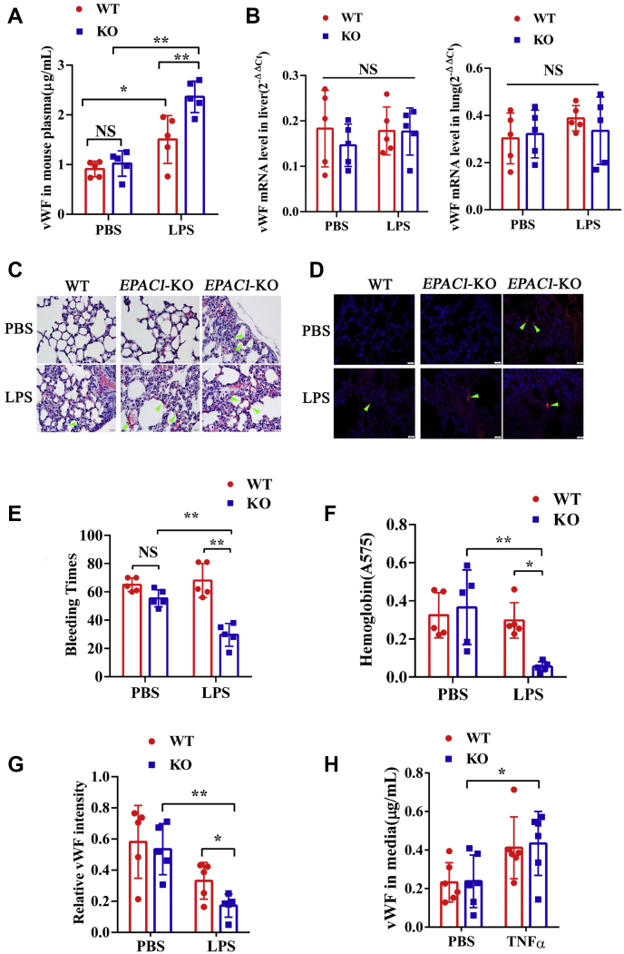
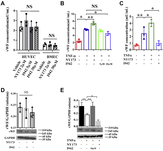
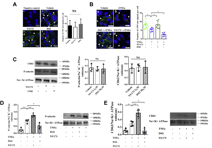
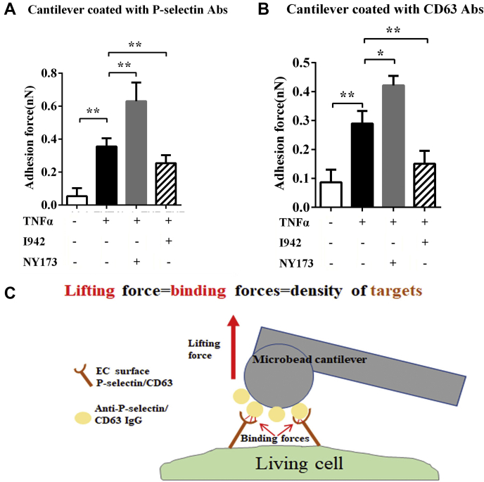
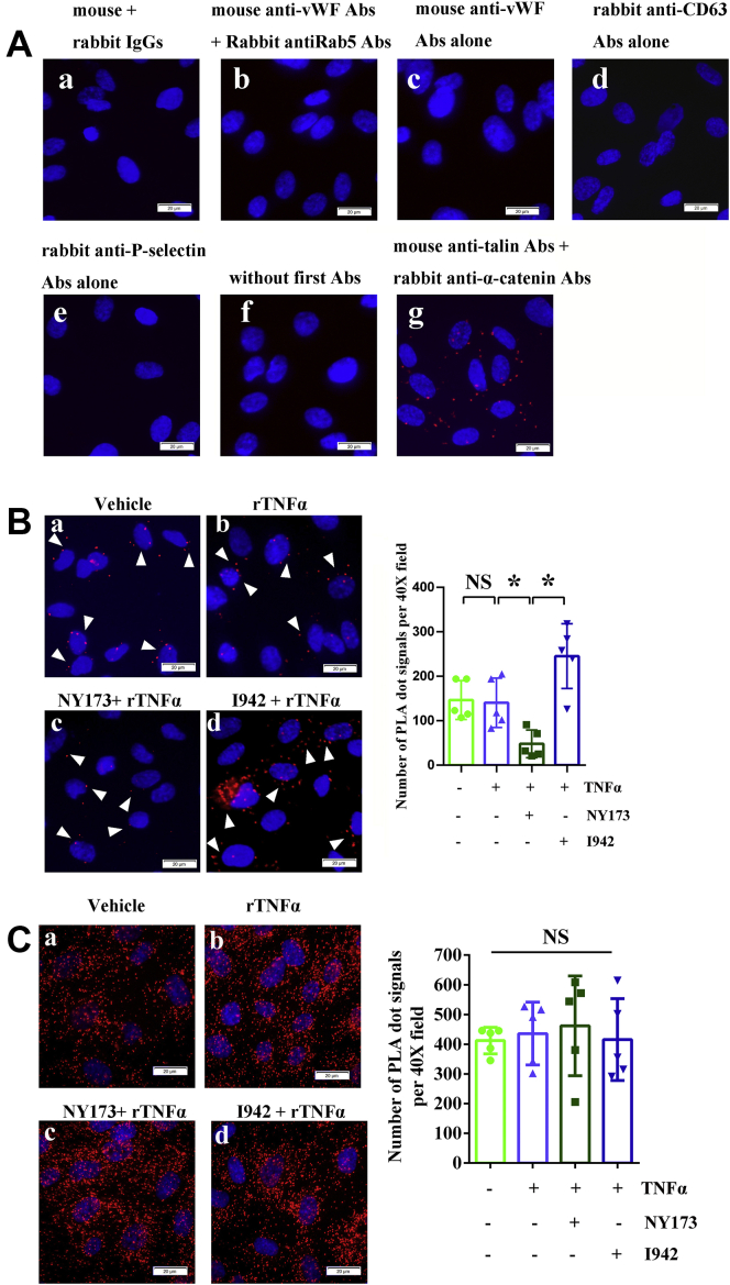
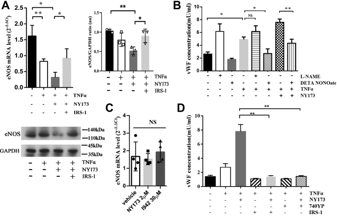
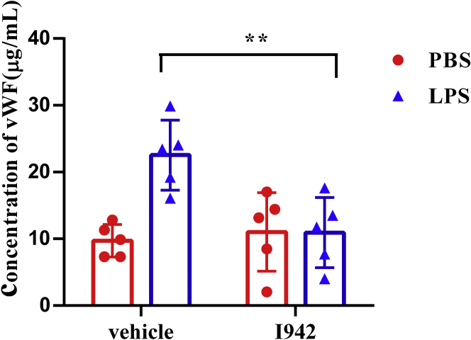
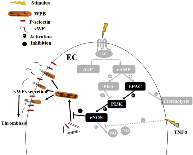
Coagulopathy is associated with both inflammation and infection, including infections with novel severe acute respiratory syndrome coronavirus-2, the causative agent Coagulopathy is associated with both inflammation and infection, including infection with novel severe acute respiratory syndrome coronavirus-2, the causative agent of COVID-19. Clot formation is promoted via cAMP-mediated secretion of von Willebrand factor (vWF), which fine-tunes the process of hemostasis. The exchange protein directly activated by cAMP (EPAC) is a ubiquitously expressed intracellular cAMP receptor that plays a regulatory role in suppressing inflammation. To assess whether EPAC could regulate vWF release during inflammation, we utilized our EPAC1-null mouse model and revealed increased secretion of vWF in endotoxemic mice in the absence of the EPAC1 gene. Pharmacological inhibition of EPAC1 in vitro mimicked the EPAC1-/- phenotype. In addition, EPAC1 regulated tumor necrosis factor-α-triggered vWF secretion from human umbilical vein endothelial cells in a manner dependent upon inflammatory effector molecules PI3K and endothelial nitric oxide synthase. Furthermore, EPAC1 activation reduced inflammation-triggered vWF release, both in vivo and in vitro. Our data delineate a novel regulatory role for EPAC1 in vWF secretion and shed light on the potential development of new strategies to control thrombosis during inflammation.

Keywords: AFM; EPAC; PI3K; Weibel–Palade body; eNOS; endothelial cell; inflammation; spatial proximity; von Willebrand factor secretion.


Conflict of interest statement

Conflict of interest The authors declare that they have no conflicts of interest with the contents of this article.

Figures

Figure 1
Figure 1
Inactivation of EPAC1 increases vWF secretion and enhances microthrombosis during inflammation. Plasma vWF concentrations and vWF mRNA expression in the tissues of WT and EPAC1-KO mice treated with or without lipopolysaccharides (LPS). A, the plasma levels of vWF between the WT and EPAC1-KO mice in the presence or absence of LPS (5 mg/kg/d, i.p) for 2 h. n = 5 for each group. B, qRT-PCR analysis of vWF mRNA expression in the lungs and livers of WT and EPAC1-KO mice treated with or without LPS. Two-way ANOVAs showed that the differences in vWF mRNA expression was not significant. n = 5 for each group. C and D, the formation of microthrombi in the microvessels of LPS-treated EPAC1-KO mice. WT (A and B) and EPAC1-KO (C–F) mice were treated with PBS (A and C) or LPS (5 mg/kg/d, i.p. ×1). (B, D–F). After 24 h, the lungs were dissected after euthanasia and whole-animal perfusion. The lungs were immersion-fixed in 10% buffered formalin overnight. Microthrombi were detected in all LPS-treated EPAC1-KO mice (D–F). The scale bars indicate 20 μm. D, IF of fibrin(ogen) were performed in the lungs. E, the bleeding time of the WT and EPAC1-KO mice treated with LPS. The LPS-treated EPAC1-KO mice have decreased tail bleeding times. F, blood loss quantified as the amount of hemoglobin (absorbance at 575 nm) released during the tail bleeding test in the LPS-treated WT and EPAC1-KO mice, ∗p < 0.05. n = 5 for each group (E and F). G, the relative intensity of immunofluorescence signals of vWF in lung sections from WT versus EPAC1-KO mice, which were thoroughly perfused with PBS and analyzed using ImageJ software. The data are expressed as the ratios of immunofluorescence intensities of vWF normalized against DAPI signals (55). n = 5 for each group. H, the vWF concentrations in the culture medium of WT versus EPAC1-KO mouse aortic ECs. n = 6 mice for each group. ∗p < 0.05 and ∗∗p < 0.01. The scale bars represent 20 μm. DAPI, 4′,6-diamidino-2-phenylindole; ECs, endothelial cells; EPAC, exchange protein directly activated by cAMP; IF, immunofluorescence; LPS, lipopolysaccharides; ns, not significant; qRT-PCR, reverse transcription quantitative PCR; TNFα, tumor necrosis factor-α; vWF, von Willebrand factor.
Figure 2
Figure 2
Pharmacological manipulations of EPAC1 modulate the inflammation-triggered vWF secretion from HUVECs. A, the effect of only NY173 or I942 on vWF secretion. HUVECs and BMECs were incubated with I942 (5 or 30 μM) or NY173 (2 μM) for 24 h. The vWF concentrations in the culture medium from ECs were detected using the human vWF ELISA kit. n = 3 for each group. B, the vWF concentrations in the culture medium of HUVECs. NY173 enhanced the secretion of vWF from rTNFα-treated HUVECs. The EPAC1-specific agonist I942 at 30 μM, but not 5 μM, significantly reduced the secretion of vWF from rTNFα-treated HUVECs. C, the vWF concentrations in the culture medium of BMECs. D, the vWF protein levels in the cell lysates of HUVECs incubated with I942 (30 μM) or NY173 (2 μM) for 24 h. E, the vWF protein levels in the cell lysates of HUVECs. vWF expression was significantly reduced in rTNFα-treated HUVECs. The vWF protein expression was further significantly decreased in the NY173 + rTNFα group. I942 (30 μM) significantly increased vWF expression in rTNFα-treated HUVECs. n = 3 for each group. ∗p < 0.05 and ∗∗p < 0.01. au, arbitrary units of the ratio of vWF to GAPDH; BMECs, brain microvascular ECs; ECs, endothelial cells; EPAC, exchange protein directly activated by cAMP; HUVECs, human umbilical vein endothelial cells; NS, not significant; TNFα, tumor necrosis factor α; rTNFα, recombinant TNFα; vWF, von Willebrand factor.
Figure 3
Figure 3
EPAC1 modulates the exocytosis of WPBs during inflammation.A, immunofluorescence of vWF in HUVECs in the noninflammation state: (a) negative control where normal mouse IgGs were used as primary antibodies, (b) vehicle-only treated cells, (c) HUVECs treated with 2 μM NY173 for 24 h, and (d) HUVECs treated with 30 μM I942 for 24 h. In b, c, and d, the samples were incubated with the anti-vWF mouse monoclonal antibody as the primary antibody and then stained with DAPI (blue) and Alexa Fluor 488–conjugated secondary antibody for vWF labeling (green). The scale bars indicate 20 μm. The quantitative analyses of vWF-positive puncta and cell nuclei were performed using ImageJ software. The results are expressed as dot signals enumerated in each cell. Five microscopic fields were examined for each sample. The results are expressed as dot signals enumerated in each cell. n  =  3 for each group. B, immunofluorescence of vWF in HUVECs in the inflammation state. The HUVECs were pretreated with NY173 or I942 for 24 h and treated with rTNFα (50 ng/ml) for 4 h: (a) vehicle only, (b) rTNFα only, (c) I942 + rTNFα, (d) NY173 + rTNFα. The scale bars represent 20 μm. ImageJ was used to quantify vWF-positive puncta. Five microscopic fields were examined for each case. The results were expressed as dot signals enumerated in each cell. n = 6 for each group (A and B). C, Western immunoblotting was performed to analyze the expression of CD63, P-selectin, and the sodium–potassium pump in the membrane fractions of HUVECs. The graphs of the relative ratios in arbitrary units (au) of P-selectin or CD63 with Na+/K+-ATPase are shown. n = 3 for each group. D, Western immunoblotting analysis of the expression of P-selectin in the membrane fractions of HUVECs treated with rTNFα only, NY173 or I942, and rTNFα, and untreated. A graph of the relative ratio in au of P-selectin with Na+/K+-ATPase is shown. E, Western immunoblotting analysis of the expression of CD63 in the membrane fractions of HUVECs treated with NY173 or I942 alone with rTNFα. A graph of the relative ratio in au of CD63 with Na+/K+-ATPase is shown. n = 4 for each group (D and E). ∗p < 0.05 and ∗∗p < 0.01. EPAC1, exchange protein directly activated by cAMP; DAPI, 4′,6-diamidino-2-phenylindole; HUVECs, human umbilical vein endothelial cells; IgG, immunoglobulin G; NS, not significant; rTNFα, recombinant TNFα; TNFα, tumor necrosis factor; WPBs, Weibel–Palade bodies; vWF, von Willebrand factor.
Figure 4
Figure 4
EPAC1 affects P-selection and CD63 residing on endothelial apical surfaces. Quantification of P-selectin (A) and CD63 (B) expression on the surface of live HUVECs as measured by atomic force microscopy (AFM). A normal mouse IgG-coated colloidal cantilever was used during calibration of the AFM system before measuring the unbinding forces between an anti-P-selectin– or anti-CD63 antibody–coated cantilever and the cell (for details, see Experimental procedures). With either anti-P-selectin– or anti-CD63 antibody–coated cantilevers, the adhesion forces were significantly stronger in the NY173 + TNFα–treated HUVECs than the TNFα-only–treated groups; ∗p < 0.05 and ∗∗p < 0.01. n = 12 for each group. C, a schematic depicting the specific unbinding force measurements that can be compiled for quantification of total P-selectin or CD63 expression on the surface areas of a live HUVEC. AFM, atomic force microscopy; EC, endothelial cell; HUVECs, human umbilical vein endothelial cells; IgG, immunoglobulin G; TNFα, tumor necrosis factor-α.
Figure 5
Figure 5
PLA signals of vWF-CD63 and vWF-P-selectin in HUVECs. A, control groups: (a) reagent-negative control where normal mouse and rabbit IgGs were used as primary Abs, (b) negative control where mouse anti-vWF Abs and rabbit anti-Rab5 Abs were used as primary Abs, (c) mouse anti-vWF Abs alone, (d) rabbit anti-CD63 Abs alone, (e) rabbit anti-P-selectin Abs alone, (f) no first Abs, and (g) positive control where mouse anti-talin Abs and rabbit anti-α-catenin were used as primary Abs (red). B and C, IFA staining: the representative PLA signals (red) of vWF-P-selectin (B, arrowheads) and vWF-CD63 (C), respectively, in HUVECs. (a) vehicle only, (b) rTNFα, (c) NY173 pretreatment + rTNFα, and (d) I942 pretreatment + rTNFα. The nuclei in panels A–C were counterstained with DAPI (blue). The scale bars indicate 20 μm. The graphs show the quantitative analysis of vWF-P-selectin PLA signals (B) and vWF-CD63 PLA signals (C) using ImageJ software. Five 40× microscopic fields were examined for each case. The results are expressed as dot signals enumerated in each 40× field. n  = 5 per group. ∗p < 0.05. Abs, antibodies; DAPI, 4′,6-diamidino-2-phenylindole; HUVECs, human umbilical vein endothelial cells; IFA, immunofluorescence assay; IgG, immunoglobulin G; NS, not significant; PLA, proximity ligation assay; rTNFα, recombinant tumor necrosis factor-α; vWF, von Willebrand factor.
Figure 6
Figure 6
EPAC regulates the vWF secretion by HUVECs in a PI3K-/eNOS-dependent manner. A, RT-qPCR analysis of eNOS mRNA expression in HUVECs and a representative protein immunoblot of eNOS. The eNOS mRNA expression decreased in the HUVECs treated with rTNFα for 4 h; in the presence of NY173 for 24 h and rTNFα for 4 h, eNOS mRNA expression further declined. The PI3K activator IRS-1 (with rTNFα treatment) reversed the effect of NY173 on mRNA expression. The immunoblot displayed that in the presence of NY173 and rTNFα, eNOS expression was suppressed, whereas adding IRS-1 reversed the effect of NY173. B, the assessment of eNOS on vWF secretion triggered by rTNFα as measured by ELISA. The data show that DETA NONOate significantly downregulated rTNFα-induced vWF secretion. The regulatory role of EPAC on rTNFα-triggered vWF secretion was eNOS-dependent. C, the effect of NY173 and I942 on eNOS mRNA expression in HUVECs, without exposure to TNFα. The HUVECs were incubated with 2 μM NY173 or 30 μM I942 for 24 h. D, the assessment by ELISA of the PI3K effect on EPAC-regulated vWF secretion by HUVECs. The vWF concentrations in culture media were detected in all groups: vehicle-only, rTNFα-only, rTNFα + NY173, rTNFα + IRIS-1, rTNFα + NY173 + IRIS-1, rTNFα +740YP, and r-TNFα + NY173 + 740YP–pretreated HUVECs. n = 3 for each group. ∗p < 0.05 and ∗∗p < 0.01. eNOS, endothelial nitric oxide synthase; EPAC, exchange protein directly activated by cAMP; HUVECs, human umbilical vein endothelial cells; NS, not significant; qRT-PCR, reverse transcription quantitative PCR; rTNFα, recombinant TNFα; TNFα, tumor necrosis factor-α; vWF, von Willebrand factor.
Figure 7
Figure 7
EPAC1-specific agonist I942 reduces LPS-induced vWF secretion in the WT mice. ELISAs were performed to analyze the plasma vWF concentrations in the C57BL/6 mice that were treated with I942. I942 was administrated at 5 mg/kg/day intraperitoneally before the mouse was exposed to LPS (5 mg/kg, i.p.) or equal volume of PBS for 2 h. I942 downregulated the plasma vWF levels in endotoxemic mice, ∗∗p < 0.01. n = 5 for each group. EPAC, exchange protein directly activated by cAMP; LPS, lipopolysaccharides; vWF, von Willebrand factor.
Figure 8
Figure 8
Model for EPAC1 regulating inflammation-triggered vWF release in a PI3K-/eNOS-dependent manner. Our proposed pathways from the present study are in black, and previously reported pathways are in gray. EC, endothelial cell; eNOS, endothelial nitric oxide synthase; EPAC, exchange protein directly activated by cAMP; NO, nitric oxide; TNFα, tumor necrosis factor-α; WPB, Weibel–Palade body; vWF, von Willebrand factor.

Update of

Similar articles

Cited by

References

    1. Ackermann M., Verleden S.E., Kuehnel M., Haverich A., Welte T., Laenger F., Vanstapel A., Werlein C., Stark H., Tzankov A., Li W.W., Li V.W., Mentzer S.J., Jonigk D. Pulmonary vascular endothelialitis, thrombosis, and angiogenesis in Covid-19. N. Engl. J. Med. 2020;383:120–128. - PMC - PubMed
    1. Connors J.M., Levy J.H. COVID-19 and its implications for thrombosis and anticoagulation. Blood. 2020;135:2033–2040. - PMC - PubMed
    1. Connors J.M., Levy J.H. Thromboinflammation and the hypercoagulability of COVID-19. J. Thromb. Haemost. 2020;18:1559–1561. - PMC - PubMed
    1. Khodamoradi Z., Boogar S.S., Shirazi F.K.H., Kouhi P. COVID-19 and acute pulmonary embolism in postpartum patient. Emerg. Infect. Dis. 2020;26:1937–1939. - PMC - PubMed
    1. Bompard F., Monnier H., Saab I., Tordjman M., Abdoul H., Fournier L., Sanchez O., Lorut C., Chassagnon G., Revel M.P. Pulmonary embolism in patients with Covid-19 pneumonia. Eur. Respir. J. 2020;56 - PMC - PubMed

MeSH terms

Substances

LinkOut - more resources