. 2021 Nov;297(5):101318.
doi: 10.1016/j.jbc.2021.101318. Epub 2021 Oct 20.

Structural determinants of the integrin transmembrane domain required for bidirectional signal transmission across the cell membrane

Affiliations

Structural determinants of the integrin transmembrane domain required for bidirectional signal transmission across the cell membrane

Zhengli Wang et al. J Biol Chem. 2021 Nov.

Abstract

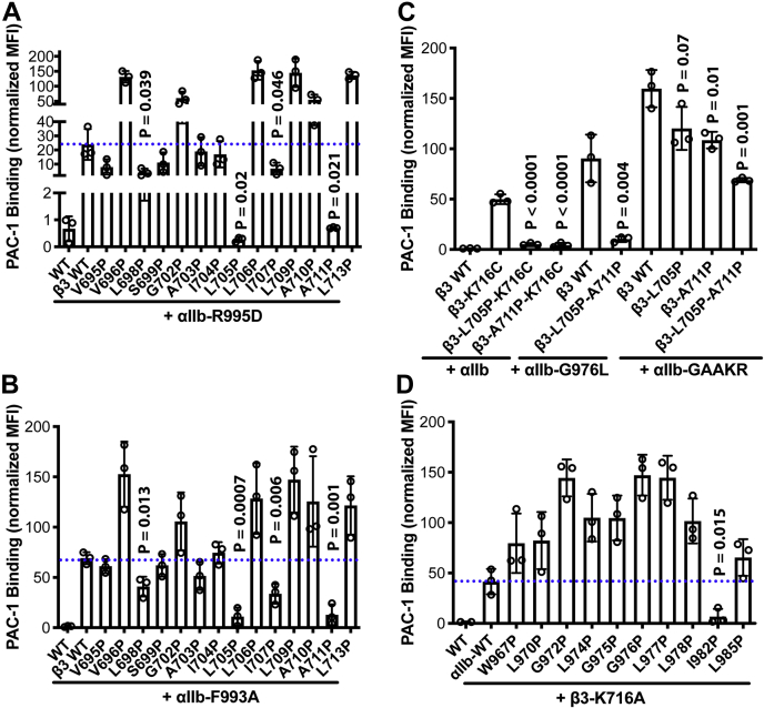
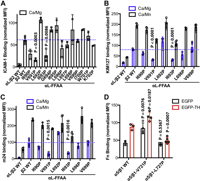
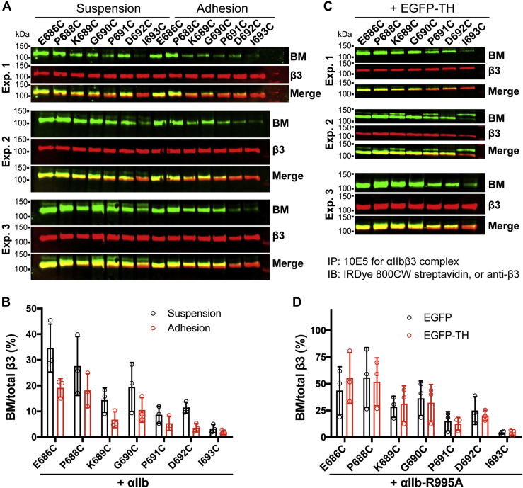
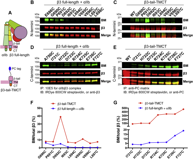
Studying the tight activity regulation of platelet-specific integrin αIIbβ3 is foundational and paramount to our understanding of integrin structure and activation. αIIbβ3 is essential for the aggregation and adhesion function of platelets in hemostasis and thrombosis. Structural and mutagenesis studies have previously revealed the critical role of αIIbβ3 transmembrane (TM) association in maintaining the inactive state. Gain-of-function TM mutations were identified and shown to destabilize the TM association leading to integrin activation. Studies using isolated TM peptides have suggested an altered membrane embedding of the β3 TM α-helix coupled with αIIbβ3 activation. However, controversies remain as to whether and how the TM α-helices change their topologies in the context of full-length integrin in native cell membrane. In this study, we utilized proline scanning mutagenesis and cysteine scanning accessibility assays to analyze the structure and function correlation of the αIIbβ3 TM domain. Our identification of loss-of-function proline mutations in the TM domain suggests the requirement of a continuous TM α-helical structure in transmitting activation signals bidirectionally across the cell membrane, characterized by the inside-out activation for ligand binding and the outside-in signaling for cell spreading. Similar results were found for αLβ2 and α5β1 TM domains, suggesting a generalizable mechanism. We also detected a topology change of β3 TM α-helix within the cell membrane, but only under conditions of cell adhesion and the absence of αIIb association. Our data demonstrate the importance of studying the structure and function of the integrin TM domain in the native cell membrane.

Keywords: cell adhesion; integrin; platelet; talin; transmembrane domain.


Conflict of interest statement

Conflict of interest Authors declare that they have no competing interests.

Figures

Figure 1
Figure 1
Proline scanning of αIIbβ3TM domains.A, sequence alignment of human integrin transmembrane domains. The predicted TM C-terminal boundary is marked with a red dashed line. The conserved small amino acids are highlighted in yellow. Shown in red are the residues that inhibit integrin inside-out activation when mutated to proline. B, backbone structure of αIIbβ3 TM domain. Backbone hydrogen bonds are shown as dashed lines in yellow. Selected residues are marked as spheres of Cα atoms. C and D, ligand mimetic PAC-1 binding to αIIbβ3 integrin with indicated proline substitutions. HEK293FT cells were transfected with indicated αIIbβ3 constructs. The ligand-mimetic mAb PAC-1 binding was performed in the buffer containing 1 mM Ca2+/Mg2+ and detected by flow cytometry. The level of PAC-1 binding was normalized to αIIbβ3 surface expression detected by mAb AP3 and shown as mean ± SD (n = 3). Unpaired two-tailed t test was performed between the control group without proline mutation and the group with proline mutation. Only p values less than 0.05 are shown. E, structural illustration of αIIbβ3 TM domain within the cell membrane in the absence and presence of selected proline mutations. The proline mutations were introduced in silico to the TM structure using PyMOL. The proline-induced broken of a rigid α-helical structure was indicated. The interfacial residues are shown as sticks or Cα spheres. MP, membrane proximal.
Figure 2
Figure 2
Proline scanning of αIIbβ3TM domains identified proline mutations that hindered activation.A and B, PAC-1 binding of β3 TM proline mutations on the background of the αIIb cytoplasmic mutations, αIIb-R995D or αIIb-F993A that mimics inside-out activation of αIIbβ3 integrin. C, PAC-1 binding of αIIb TM proline mutations on the background of the β3 cytoplasmic mutations, β3-F993A that mimics inside-out activation of αIIbβ3 integrin. D, PAC-1 binding of selected β3 TM proline mutations on the background of the αIIb mutations, αIIb-G976L or αIIb-GAAKR that mimics inside-out activation of αIIbβ3 integrin. HEK293FT cells were transfected with indicated αIIbβ3 constructs. The PAC-1 binding was performed in the buffer containing 1 mM Ca2+/Mg2+ and detected by flow cytometry. The level of PAC-1 binding was normalized to αIIbβ3 surface expression detected by mAb AP3 and shown as mean ± SD (n = 3). Unpaired two-tailed t test was performed between the control group without proline mutation and the group with proline mutation.
Figure 3
Figure 3
Effect of disturbing the rigidity of TM and CT domains on talin1-head induced αIIbβ3activation.A, effect of the selected αIIbβ3 TM proline mutations on EGFP-talin-head (EGFP-TH) induced PAC-1 binding to αIIbβ3 integrin. B, the β3-GS or β3-GGGS mutation was generated by the insertion of GS or GGGS sequence before the conserved β3-D723 residue. C, effect of β3-GS and β3-GGGS mutations on EGFP-TH induced PAC-1 binding to αIIbβ3 integrin. HEK293FT cells were transfected with indicated αIIbβ3 plus EGFP or EGFP-TH constructs. The PAC-1 binding was performed in the buffer containing 1 mM Ca2+/Mg2+ and detected by flow cytometry. The PAC-1 binding of EGFP and αIIbβ3 double-positive cells was normalized to αIIbβ3 surface expression detected by mAb AP3 and shown as mean ± SD (n ≥ 3). Unpaired two-tailed t test was performed between the control group without proline mutation and the group with proline mutation. D, interaction of β3-GS and β3-GGGS mutants with talin1-head domain. HEK293FT cells were transfected with αIIb WT and indicated β3 plus EGFP-TH constructs. Total cell lysates were immunoprecipitated with anti-EGFP antibody and immunoblotted with anti-β3 antibody.
Figure 4
Figure 4
Effect of TM proline mutations on the activation of β2and β1integrins.A, effect of TM proline mutations of β2 integrin on ICAM-1 binding to αLβ2 integrin. The ICAM-1 binding to αLβ2-transfected HEK293FT cells was performed in the presence of 1 mM CaCl2 plus 1 mM MgCl2 (Ca/Mg). B and C, effect of TM proline mutations of β2 integrin on the binding of activation-dependent mAbs, KIM127 and m24. The mAb binding to αLβ2-transfected HEK293FT cells was performed in the presence of 1 mM Ca/Mg or 1 mM CaCl2 plus 1 mM MnCl2 (Ca/Mn). D, effect of TM proline mutations of β1 integrin on talin-head (TH)-induced fibronectin (Fn) binding to α5β1 integrin. α5β1-KO HEK293FT cells were transfected with α5β1 plus EGFP or EGFP-TH constructs. The binding of Alexa Fluor 647-labled human fibronectin (Fn) was performed in the presence of 1 mM Ca/Mg. Both the ligand and the LIBS mAb binding were normalized to integrin expression. Data are mean ± SD (n ≥ 3). Unpaired two-tailed t test was performed between the control group without proline mutation and the group with proline mutation.
Figure 5
Figure 5
Effect of TM proline mutations on αIIbβ3-mediated cell spreading.AC, HEK293 cells stably expressing αIIb3 WT, αIIb3-L705P-A711P, or αIIb-I982P/β3 spread on immobilized PAC-1. D and E, HEK293 cells stably expressing αIIb3 WT or αIIb3-L705P-A711P spread on immobilized fibrinogen. Cells were allowed to adhere on platelets coated with 5 μg/ml PAC-1 or 25 μg/ml fibrinogen at 37 °C for 1 h and then fixed and stained with Alexa-Fluor-488-labeled AP3 for β3 detection and Alexa-Fluor-564-labeled phalloidin for F-actin detection. Nuclei were stained with DAPI in panels AC. F, quantification of cell spreading areas in panels AE. The averaged cell areas were calculated based on 30 to 50 cells for each of three independent repeats. Data are mean ± SD. Unpaired two-tailed t test was performed between the WT and the mutant cells.
Figure 6
Figure 6
BM-labeling of αIIbβ3TM cysteine substitutions before and after soluble ligand binding.A, the cysteine scanning accessibility method for determining the burial/exposure status of integrin TM domain. A single cysteine mutation was introduced into the α or β TM domain. The cysteine located outside of the cell membrane can be labeled by biotin-maleimide (BM), while the cysteine buried in the cell membrane is not accessible to the labeling. BE, the BM labeling of substituted cysteines in the β3 (B and D) and αIIb (C and E) TM domains. HEK293FT cells were transfected with wild-type αIIb plus β3 containing a single-cysteine mutation or wild-type β3 plus αIIb containing a single-cysteine mutation. The cells were treated with or without the high-affinity ligand eptifibatide (Ept) and labeled with BM on ice. The αIIbβ3 was immunoprecipitated with αIIbβ3-complex specific mAb 10E5 and immunoblotted with indicated detection reagents. FI, quantification of western blot data as described in panels BE. Data are mean ± SD (n ≥ 2).
Figure 7
Figure 7
BM labeling of the cysteine substitutions in the membrane-proximal (MP) region of αIIbcytoplasmic tail.A, the introduction of a disulfide bond that cross-links the αIIb and β3 subunit at the N-terminus of TM domain. HEK293FT cells were transfected with β3 WT or β3-P688C mutant and the αIIb MP cysteine mutants in the presence of L959C mutation. The αIIbβ3 proteins were immunoprecipitated with mAb 10E5 and subjected to immunoblot with anti-β3 mAb AP3 under nonreduced condition. B, disulfide bond formation between αIIb-L959C and β3-P688C reversed the αIIbβ3 activation induced by the cysteine substitutions at the αIIb MP region. HEK293FT cells were transfected with indicated αIIb constructs plus β3-WT or β3-P688C mutant. PAC-1 binding to the transfected cells was performed in the presence of 1 mM Ca/Mg. Data are mean ± SD (n ≥ 2). C, BM labeling of αIIb MP cysteine mutants in the absence and presence of the disulfide bond formed between αIIb-L959C and β3-P688C. D, quantification of western blot data as described in panel C. Data are mean ± SD (n ≥ 2).
Figure 8
Figure 8
BM labeling of the cysteine substitutions in the β3TM domain upon cell adhesion or EGFP-TH-induced αIIbβ3activation.A, HEK293FT cells were transfected with αIIb WT plus indicated β3 cysteine mutants. The transfected cells were kept in suspension or allowed to adhere on fibrinogen-coated surface and labeled with BM. B, quantification of western blot data of panel A. C, HEK293FT cells were transfected with EGFP-TH and αIIb-R995A plus indicated β3 cysteine mutants. The cells were labeled with BM in suspension. D, quantification of western blot data of panel C. Data are mean ± SD of three independent experiments.
Figure 9
Figure 9
BM labeling of the cysteine substitutions in the β3TM domain that was fully separated from the αIIbsubunit.A, design of β3-tail-TMCT construct. A protein C tag was added to the N-terminus of β3-tail-TMCT construct. B and D, BM labeling of the cysteine substitutions in the TM domain of full-length β3 coexpressed with αIIb in HEK293FT cells. C and E, BM labeling of the cysteine substitutions in the TM domain of β3-tail-TMCT expressed in HEK293FT cells. F, quantification of western blot data of panels B and C. G, quantification of western blot data of panels D and E. Data are presented as BM signal as a percentage of total β3 signal.

Similar articles

Cited by

References

    1. Hynes R.O. Integrins: Bi-directional, allosteric, signalling machines. Cell. 2002;110:673–687. - PubMed
    1. Springer T.A., Dustin M.L. Integrin inside-out signaling and the immunological synapse. Curr. Opin. Cell Biol. 2012;24:107–115. - PMC - PubMed
    1. Kechagia J.Z., Ivaska J., Roca-Cusachs P. Integrins as biomechanical sensors of the microenvironment. Nat. Rev. Mol. Cell Biol. 2019;20:457–473. - PubMed
    1. Kim C., Ye F., Ginsberg M.H. Regulation of integrin activation. Annu. Rev. Cell Dev. Biol. 2011;27:321–345. - PubMed
    1. Coller B.S. alphaIIbbeta3: Structure and function. J. Thromb. Haemost. 2015;13 Suppl 1:S17–25. - PMC - PubMed

Publication types

MeSH terms

Substances

LinkOut - more resources