. 2021 Nov;297(5):101316.
doi: 10.1016/j.jbc.2021.101316. Epub 2021 Oct 20.

Progesterone receptor membrane component 1 (PGRMC1) binds and stabilizes cytochromes P450 through a heme-independent mechanism

Affiliations

Progesterone receptor membrane component 1 (PGRMC1) binds and stabilizes cytochromes P450 through a heme-independent mechanism

Meredith R McGuire et al. J Biol Chem. 2021 Nov.

Abstract

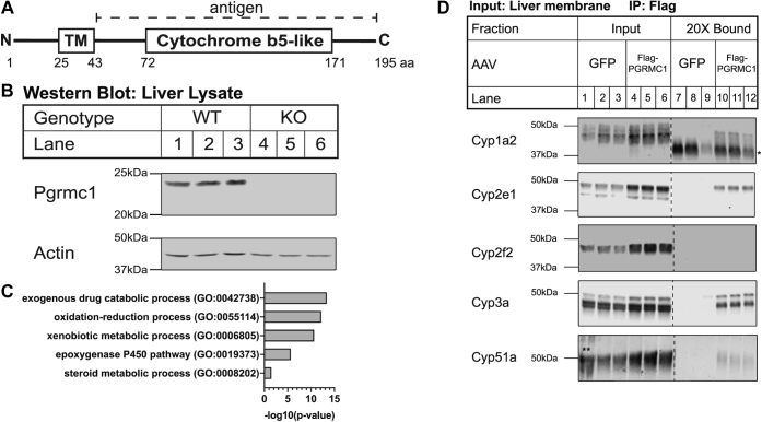
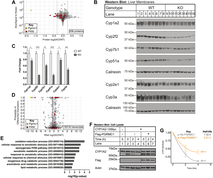
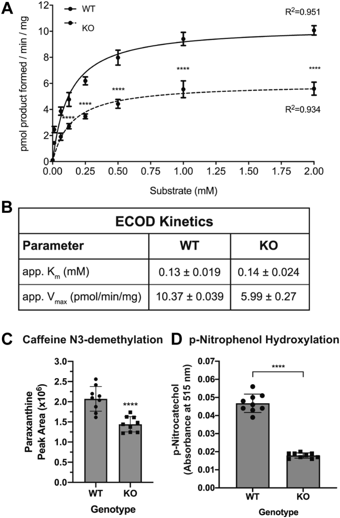
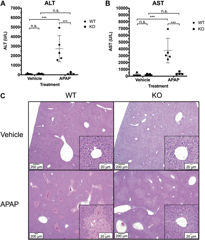
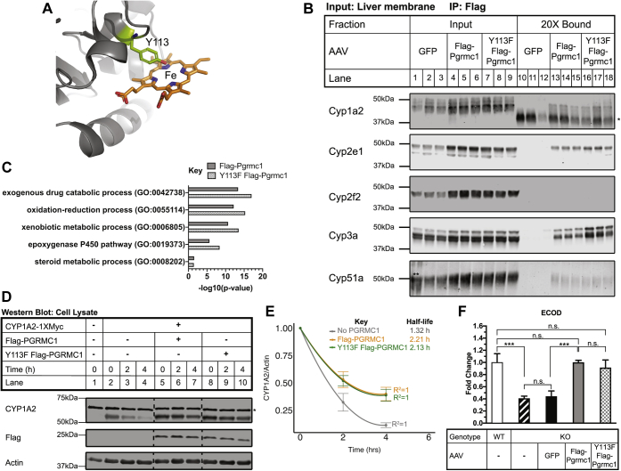
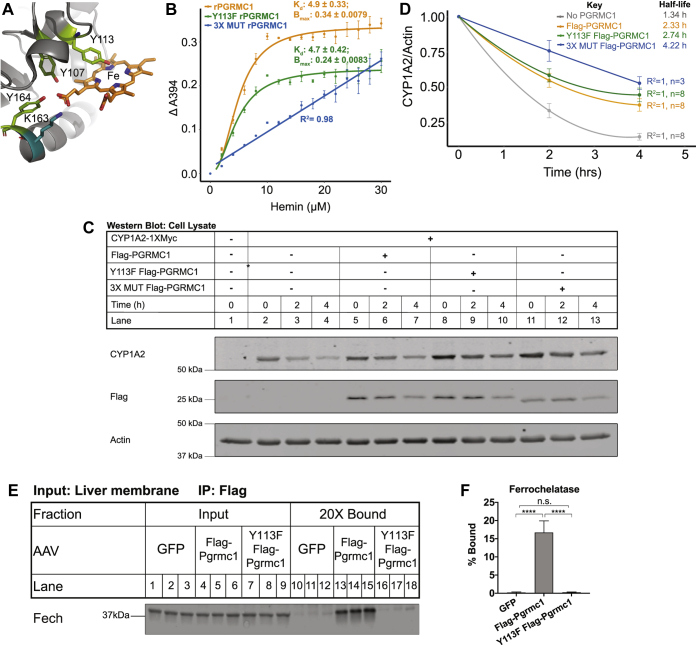
Progesterone receptor membrane component 1 (PGRMC1) is a heme-binding protein implicated in a wide range of cellular functions. We previously showed that PGRMC1 binds to cytochromes P450 in yeast and mammalian cells and supports their activity. Recently, the paralog PGRMC2 was shown to function as a heme chaperone. The extent of PGRMC1 function in cytochrome P450 biology and whether PGRMC1 is also a heme chaperone are unknown. Here, we examined the function of Pgrmc1 in mouse liver using a knockout model and found that Pgrmc1 binds and stabilizes a broad range of cytochromes P450 in a heme-independent manner. Proteomic and transcriptomic studies demonstrated that Pgrmc1 binds more than 13 cytochromes P450 and supports maintenance of cytochrome P450 protein levels posttranscriptionally. In vitro assays confirmed that Pgrmc1 KO livers exhibit reduced cytochrome P450 activity consistent with reduced enzyme levels. Mechanistic studies in cultured cells demonstrated that PGRMC1 stabilizes cytochromes P450 and that binding and stabilization do not require PGRMC1 binding to heme. Importantly, Pgrmc1-dependent stabilization of cytochromes P450 is physiologically relevant, as Pgrmc1 deletion protected mice from acetaminophen-induced liver injury. Finally, evaluation of Y113F mutant Pgrmc1, which lacks the axial heme iron-coordinating hydroxyl group, revealed that proper iron coordination is not required for heme binding, but is required for binding to ferrochelatase, the final enzyme in heme biosynthesis. PGRMC1 was recently identified as the causative mutation in X-linked isolated pediatric cataract formation. Together, these results demonstrate a heme-independent function for PGRMC1 in cytochrome P450 stability that may underlie clinical phenotypes.

Keywords: cytochrome P450; drug metabolism; enzyme degradation; heme; liver metabolism; protein turnover.


Figures

Figure 1
Figure 1
Pgrmc1 binds cytochromes P450 in mouse liver.A, schematic of human PGRMC1 protein (Uniprot O00264). The 195 amino acid protein consists of a single-pass transmembrane domain (TM) and cytochrome b5-like domain, which shares 30% identity with the human cytochrome b5 protein (NP_683725.1). A rabbit polyclonal antibody (5944) was raised to a bacterially expressed recombinant protein consisting of amino acids 43 to 195 of human PGRMC1. B, Pgrmc1 protein expression in Pgrmc1 KO mouse liver. Pgrmc1 was knocked out in the whole animal by crossing mice with a conditionally targeted Pgrmc1 allele containing loxP sites flanking exons 1 and 2 of the gene to Sox2-Cre mice. Knockout in the liver was confirmed by western blotting liver lysate (+β-mercaptoethanol) with an anti-PGRMC1 antibody (5944). Actin is a loading control. Each lane is a biological replicate (WT n = 3, KO n = 3). C, biological process gene ontology (GO) Term analysis on candidate binding partners of Flag-Pgrmc1 from liver. Pgrmc1 KO mice were infected with 5 × 1011 particles of AAV8 GFP or AAV8 Flag-Pgrmc1 by tail vein injection and sacrificed after 8 days. Liver membrane fractions were subjected to Flag coimmunoprecipitation. Eluates from technical triplicates were pooled for each of three biological replicates and tagged with isobaric labels. Flag-Pgrmc1-binding proteins were identified by mass spectrometry—33 proteins have a fold-change ≥20% compared with the GFP control. The GO terms enriched in relation to the complete Mus musculus proteome were identified using PANTHER. A Fisher’s exact test and Bonferroni correction were used to determine enriched GO terms with a p-value ≤ 0.05. D, input (×) and bound (20×) fractions from Flag coimmunoprecipitation samples were subjected to western blotting for cytochromes P450 detected by mass spectrometry. Each panel is a montage from a single membrane with dashed lines denoting removed lanes. (∗ denotes IgG; ∗∗ ladder overflow into Lane 1.)
Figure 2
Figure 2
Pgrmc1 regulates cytochrome P450 protein levels.A, membrane-enriched proteome of Pgrmc1 KO livers. Steady-state protein levels from WT and Pgrmc1 KO liver membrane fractions were quantified by mass spectrometry with isobaric tagging. Membrane proteins from the livers of four to five male mice of each genotype were pooled. Two biological replicates were conducted with 936 proteins measured in both replicates and further analyzed. The log2 fold-change [log2(KO/WT)] in expression was plotted against the absolute value of the signal-to-noise ratio. The signal-to-noise ratio is a moderated test statistic and reflects how unusually a given value of the log2 fold-change is when considering the whole data set. Proteins with an absolute value of the signal-to-noise ratio ≥2 were considered significant. Blue dashed lines indicate a 20% fold change, cytochromes P450 are colored red, and Pgrmc1 is colored yellow. B, Western blots of liver membrane fractions for cytochromes P450. Liver membrane-enriched protein (15 μg/lane for Cyp2f2, Cyp51a, and Cyp7b1, 10 μg/lane for all others; +β-mercaptoethanol; calnexin panels are loading controls for the panels above them) was analyzed by western blotting using the indicated antibodies. Each lane is a biological replicate (WT n = 8, KO n = 8). C, fold change in liver membrane protein expression of cytochromes P450 in Pgrmc1 KO compared with WT mice for (B). Cytochrome P450 signal intensities for each lane in (B) were first normalized to calnexin. Error bars are 1 SEM. (WT n = 8, KO n = 8; Welch’s t test, one-tailed; ∗ p ≤ 0.05, ∗∗ p ≤ 0.01, ∗∗∗ p ≤ 0.001, ∗∗∗∗ p ≤ 0.0001). D, transcriptome of Pgrmc1 KO livers. RNA-seq was performed on total RNA from Pgrmc1 KO mice and WT controls. RNAs from five mice per genotype were pooled to produce one sample per genotype for analysis. In total, 16,318 genes were measured and plotted. The log2 fold change [log2(KO/WT)] in expression was plotted against the probability of differential expression (PDE), which is the Bayesian posterior probability that a difference in expression exists. Genes with a PDE ≥0.95 were considered significant. Blue dashed lines indicate a 40% fold change, cytochromes P450 are colored red, and Pgrmc1 is colored yellow. E, biological process GO Term analysis on transcripts more abundant in Pgrmc1 KO liver. Enriched GO terms among the 70 genes with a fold-change ≥40% and PDE ≥0.95 as compared with all transcripts measured were identified using PANTHER. A Fisher’s exact test and Bonferroni correction were used to determine enriched GO terms with a p-value ≤ 0.05. F, flag-PGRMC1 stabilizes CYP1A2 in human SV589 cells. PGRMC1 KO cells were cotransfected with 5 μg CYP1A2-1XMyc and 10 μg of empty vector (pcDNA3.1) or Flag-PGRMC1 in a 10-cm plate. At 24 h posttransfection, cells were split 1:6 into a 6-well plate. At 48 h posttransfection, cells were treated with 100 μg/ml emetine and harvested every 2 h. Cell lysates were analyzed by western blotting. Actin is a loading control. Panels are representative of five independent experiments. Each panel is a montage from a single membrane with dashed lines denoting removed lanes. (∗ denotes background band). G, the half-life of CYP1A2 in the presence and absence of Flag-PGRMC1 was determined from (F). CYP1A2 signal was normalized to the actin loading control signal. Within each replicate, expression was normalized to the t = 0 value for each condition and then averaged. The data were fit to a second-order polynomial linear model (R2 WT = 1, KO = 1). The half-life is calculated as the x-coordinate of the curve when y = 0.5. Error bars are 1 SEM (No PGRMC1 n = 5, Flag-PGRMC1 n = 5).
Figure 3
Figure 3
Pgrmc1 KO mouse livers display reduced Cyp1a2 and Cyp2e1 activities.A, 7-ethoxycoumarin O-deethylation (ECOD) reaction in Pgrmc1 KO membrane fractions. Increasing concentrations of 7-ethoxycoumarin (0, 0.0625, 0.125, 0.25, 0.5, 1, 2 mM) were combined with 40 μg of membrane protein from WT or Pgrmc1 KO livers and the NADPH-dependent formation of 7-hydroxycoumarin in 30 min at 37 °C was assayed. Samples were pooled liver membrane protein from five male mice of each genotype. Enzyme kinetics of the ECOD reaction were fit to the Michaelis–Menten equation. Error bars are 1 SD (WT n = 3, KO n = 3 technical replicates; Student’s t test for each substrate concentration; ∗∗∗∗ p < 0.0001; nonlinear regression R2 WT= 0.951, KO= 0.934). B, apparent Km and Vmax of the ECOD reaction were calculated from the fitted Michaelis–Menten curves in (A). Values are mean ± SEM. C, caffeine N3-demethylation reaction in Pgrmc1 KO membrane fraction. Liver membrane protein (100 μg) from WT or Pgrmc1 KO mice prepared as in A was combined with 50 μM caffeine and an NADPH regeneration buffer system for 60 min at 37 °C. Paraxanthine formed was detected by mass spectrometry. Error bars are 1 SD (WT n = 9, KO n = 9 technical replicates; Student’s t test; ∗∗∗∗ p < 0.0001). D, p-Nitrophenol hydroxylation reaction in Pgrmc1 KO membrane fraction. Samples were pooled liver membrane protein from four male mice of each genotype. Liver membrane protein (125 μg) from WT or Pgrmc1 KO mice was combined with 100 μM p-nitrophenol and an NADPH regeneration buffer system for 60 min at 37 °C. Control samples contained no liver membrane protein. p-Nitrocatechol formed was detected spectrophotometrically and absorbances were corrected for background signal. Error bars are 1 SD (Control n = 6, WT n = 9, KO n = 9 replicates; Welch’s t test; ∗∗∗∗ p < 0.0001).
Figure 4
Figure 4
Pgrmc1 protects against acetaminophen (APAP)-induced liver injury.A and B, serum markers of liver injury in Pgrmc1 KO mice treated with APAP. After an overnight fast (16 h), mice were injected i.p. with 600 mg APAP/kg body weight in 50% DMSO/saline (v/v) and sacrificed 24 h after exposure. Serum ALT (A) and AST (B) were measured. Error bars are 1 SD (Vehicle: WT = 5 and KO = 5, APAP: WT = 5 and KO = 4; two-way ANOVA and Tukey’s HSD; “n.s.” is not significant, ∗∗∗ p ≤ 0.001). C, representative H&E stained sections of formalin fixed liver tissue from the mice in (A) and (B) showing a low magnification image of the liver section. Inset shows image around the central vein at high magnification. (Vehicle: WT = 5 and KO = 5, APAP: WT = 5 and KO = 5).
Figure 5
Figure 5
Y113F PGRMC1 binds and stabilizes cytochromes P450.A, structure of truncated human PGRMC1 with heme axial ligand Y113 highlighted (PDB 4X8Y). B, input (1×) and bound (20×) fractions from Flag coimmunoprecipitation samples were subjected to western blotting for cytochromes P450 detected by mass spectrometry. Lanes 1 to 6 and 10 to 15 are identical to images in Figure 1D Lanes 1 to 6 and 7 to 12. (∗ denotes IgG; ∗∗ denotes ladder overflow into Lane 1). C, biological process GO Term analysis on candidate binding partners of Flag-Pgrmc1 and Y113F Flag-Pgrmc1 from liver. Pgrmc1 KO mice were infected with AAV8 Y113F Flag-Pgrmc1 and processed as in Figure 1C. Y113F Flag-Pgrmc1 binding proteins were identified by mass spectrometry; 75 proteins have a fold change ≥20% compared with GFP. GO terms enriched in relation to the complete M. musculus proteome were identified using PANTHER. A Fisher’s exact test and Bonferroni correction were used to determine enriched GO terms with a p-value ≤ 0.05. D, PGRMC1 KO cells were cotransfected with 5 μg CYP1A2-1XMyc and 10 μg of empty vector, Flag-PGRMC1, or Y113F Flag-PGRMC1 in a 10-cm plate. At 24 h posttransfection, cells were split 1:6 into a 6-well plate. At 48 h posttransfection, cells were treated with 100 μg/ml emetine and harvested every 2 h. Cell lysates were analyzed by western blotting. Actin is a loading control. Panels are representative of five independent experiments. Each panel is a montage from a single membrane with dashed lines denoting removed lanes. Lanes 1 to 7 are the same images as Figure 2F Lanes 1 to 7. (∗ denotes background band). E, the half-life of CYP1A2 in the presence and absence of Flag-PGRMC1 or Y113F Flag-PGRMC1 was determined from (D). Half-lives were calculated as in Figure 2G. Error bars are 1 SEM. (No PGRMC1 n = 5, Flag-PGRMC1 n = 5, Y113F Flag-PGRMC1 n = 5). F, ECOD reaction in liver membranes of Pgrmc1 KO mice infected with AAV8 Y113F Flag-Pgrmc1. The reaction was conducted as in Figure 3A with 2 mM 7-ethoxycoumarin. Uninfected WT and KO samples were prepared as in Figure 3A. The infected KO samples were pooled samples from three male mice of each treatment group (AAV8 GFP, AAV8 Flag-Pgrmc1, AAV8 Y113F Flag-Pgrmc1) infected as in Figure 1C. Error bars are 1 SD (n = 3 technical replicates per condition; one-way ANOVA and Tukey HSD; “n.s.” is not significant, ∗∗∗ p ≤ 0.001).
Figure 6
Figure 6
Pgrmc1 binds cytochromes P450 in a heme-independent manner, while binding to ferrochelatase is sensitive to the Y113F mutation in PGRMC1.A, structure of truncated human PGRMC1 with heme ligands Y107, Y113, K163, and Y164 highlighted (PDB 4X8Y). B, heme-binding affinity of rPGRMC1, Y113F rPGRMC1, and 3X MUT rPGRMC1 protein. Each protein (10 μM) was incubated with 0 to 30 μM hemin for 16 h at room temperature. The amount of hemin bound was measured spectrophotometrically at A394. Data were fit to the Hill equation or a linear model as appropriate. C, 3X MUT Flag-PGRMC1 stabilizes CYP1A2 in human cells. PGRMC1 KO cells were cotransfected with 5 μg CYP1A2-1XMyc and 10 μg of either empty vector, Flag-PGRMC1, Y113F Flag-PGRMC1, or 3X MUT Flag-PGRMC1 in a 10-cm plate. At 24 h posttransfection, cells were split 1:6 into a 6-well plate. At 48 h posttransfection, cells were treated with 100 μg/ml emetine and harvested every 2 h. Cell lysates were analyzed by western blotting. Actin is a loading control. Panels are representative of three independent experiments. D, the half-life of CYP1A2 in the presence and absence of Flag-PGRMC1, Y113F Flag-PGRMC1, or 3X MUT Flag-PGRMC1 was determined from (C) and Figure 5D. Half-lives were calculated as in Figure 2G. Error bars are 1 SEM. (No PGRMC1 n = 8, Flag-PGRMC1 n = 8, Y113F Flag-PGRMC1 n = 8, 3X MUT Flag-PGRMC1 n = 3). E, input (1×) and bound (20×) fractions from Flag coimmunoprecipitation samples were subjected to western blotting for ferrochelatase, which was detected by mass spectrometry to bind Flag-Pgrmc1 in Pgrmc1 KO liver membranes. F, quantification of ferrochelatase in the bound fraction of the Flag coimmunoprecipitation for (E). Within each biological replicate, the ratio of cytochrome P450 expression in the Bound fraction to the Input fraction was quantified. Error is 1 SD (GFP n = 3, Flag-Pgrmc1 n = 3, Y113F Flag-Pgrmc1 n = 3); one-way ANOVA and Tukey HSD; n.s. denotes not significant, ∗∗∗∗p ≤ 0.0001).

Similar articles

Cited by

References

    1. Ortiz de Montellano P.R., editor. Cytochrome P450: Structure, Mechanism, and Biochemistry. 3rd Ed. Kluwer Academic/Plenum Publishers; New York, NY: 2005.
    1. Werck-Reichhart D., Feyereisen R. Cytochromes P450: A success story. Genome Biol. 2000;1 REVIEWS3003. - PMC - PubMed
    1. Hughes A.L., Powell D.W., Bard M., Eckstein J., Barbuch R., Link A.J., Espenshade P.J. Dap1/PGRMC1 binds and regulates cytochrome P450 enzymes. Cell Metab. 2007;5:143–149. - PubMed
    1. McCallum M.L., Pru C.A., Niikura Y., Yee S.P., Lydon J.P., Peluso J.J., Pru J.K. Conditional ablation of progesterone receptor membrane component 1 results in subfertility in the female and development of endometrial cysts. Endocrinology. 2016;157:3309–3319. - PMC - PubMed
    1. Peluso J.J., Liu X., Saunders M.M., Claffey K.P., Phoenix K. Regulation of ovarian cancer cell viability and sensitivity to cisplatin by progesterone receptor membrane component-1. J. Clin. Endocrinol. Metab. 2008;93:1592–1599. - PubMed

Publication types