. 2022 Jan;298(1):101290.
doi: 10.1016/j.jbc.2021.101290. Epub 2021 Oct 20.

Development of a highly specific and sensitive VHH-based sandwich immunoassay for the detection of the SARS-CoV-2 nucleoprotein

Affiliations

Development of a highly specific and sensitive VHH-based sandwich immunoassay for the detection of the SARS-CoV-2 nucleoprotein

Marion Gransagne et al. J Biol Chem. 2022 Jan.

Abstract

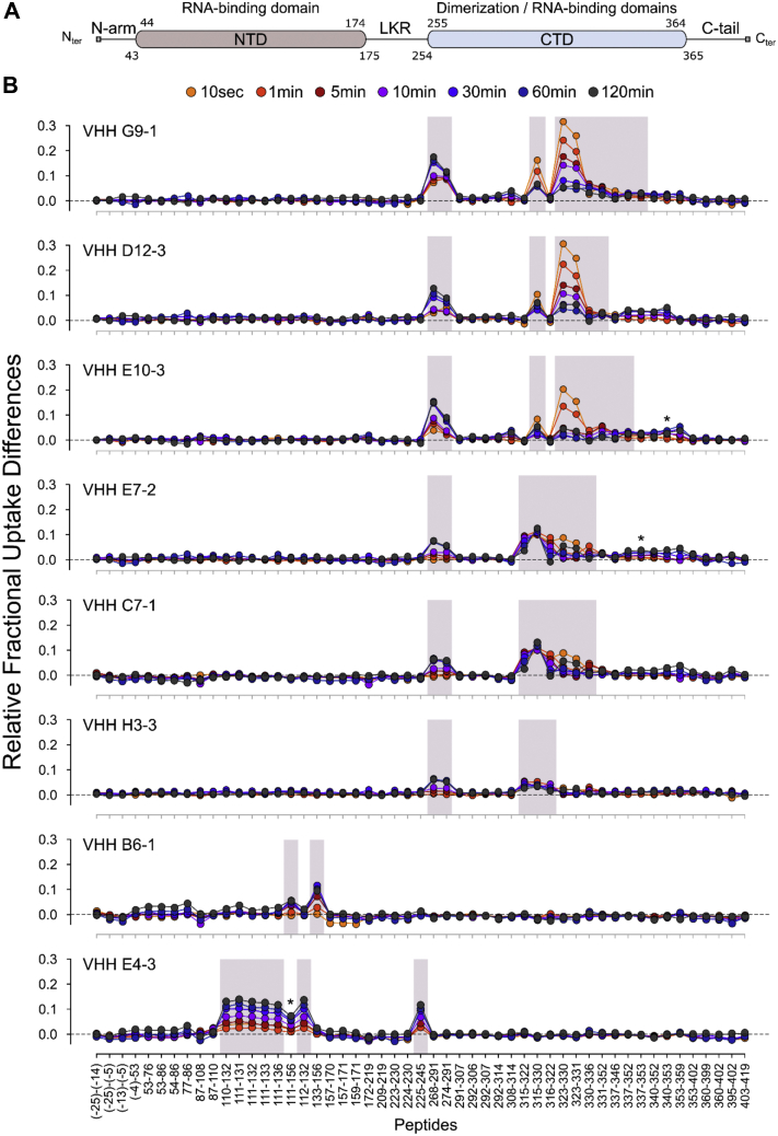
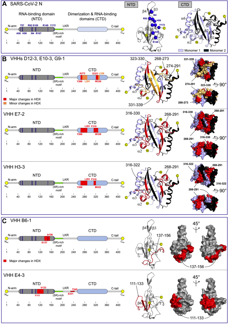
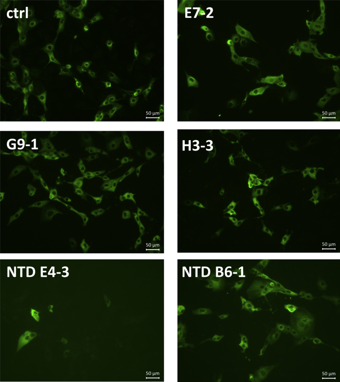
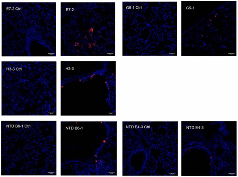
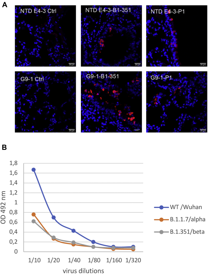
The current COVID-19 pandemic illustrates the importance of obtaining reliable methods for the rapid detection of SARS-CoV-2. A highly specific and sensitive diagnostic test able to differentiate the SARS-CoV-2 virus from common human coronaviruses is therefore needed. Coronavirus nucleoprotein (N) localizes to the cytoplasm and the nucleolus and is required for viral RNA synthesis. N is the most abundant coronavirus protein, so it is of utmost importance to develop specific antibodies for its detection. In this study, we developed a sandwich immunoassay to recognize the SARS-CoV-2 N protein. We immunized one alpaca with recombinant SARS-CoV-2 N and constructed a large single variable domain on heavy chain (VHH) antibody library. After phage display selection, seven VHHs recognizing the full N protein were identified by ELISA. These VHHs did not recognize the nucleoproteins of the four common human coronaviruses. Hydrogen Deuterium eXchange-Mass Spectrometry (HDX-MS) analysis also showed that these VHHs mainly targeted conformational epitopes in either the C-terminal or the N-terminal domains. All VHHs were able to recognize SARS-CoV-2 in infected cells or on infected hamster tissues. Moreover, the VHHs could detect the SARS variants B.1.17/alpha, B.1.351/beta, and P1/gamma. We propose that this sandwich immunoassay could be applied to specifically detect the SARS-CoV-2 N in human nasal swabs.

Keywords: COVID-19; antibody engineering; diagnostic; hydrogen-deuterium exchange; immunochemistry; nanobodies; nucleoprotein; phage display; single domain antibodies.


Conflict of interest statement

Conflict of interest The authors declare that they have no conflicts of interest with the contents of this article.

Figures

Figure 1
Figure 1
Characterization of the Nucleoprotein. A shows the SDS-PAGE gel with lanes 5–12 representing the eluted fractions containing the purified SARS-CoV-2 N and lanes 13–18 are separated contaminants; B represents the intact mass measurement. The measured molecular weight (48752.80 ± 1.96 Da) is consistent with the expected average mass calculated from the full-length SARS-CoV-2 N primary sequence (48752.13 Da, Δm = +0.67 Da (+13.7 ppm)), thereby confirming the structural integrity of the protein; C shows one main homogeneous population by DLS with a hydrodynamic radius of 6 nm. No aggregates are detectable at 37 °C; D represents the AUC measurement where 96% of the sample is under a dimeric form.
Figure 2
Figure 2
Analysis of the binding of the different VHHs to the SARS-CoV-2 Nucleoprotein. A, binding of the different VHHs to SARS-CoV-2 recombinant Nucleoprotein determined by ELISA. N was coated at 1 μg/ml and VHHs at different concentrations were then added. B, real-time monitoring of the VHH/N interaction by SPR. The determined kinetic parameters of the VHHS are provided in Table 1. C, binding of the VHHs by ELISA on cell extracts. The VHH concentration leading to maximal difference obtained between infected and uninfected cell extracts are indicated by gray variations.
Figure 3
Figure 3
Identification of the VHH binding sites by HDX-MS. A, general organization of full-length SARS-CoV-2 N showing the position of the 2-folded NTD and CTD structural domains and the three intrinsically disordered regions (N-arm, LKR, and C-tail). B, differential fractional uptake plots showing the relative variations in deuterium incorporation imposed by the binding of each VHH to full-length SARS-CoV-2 N. The differences in uptake between the apo- and VHH-bound states were calculated for each peptide and time point and plotted as a function of peptide position. A positive uptake difference is indicative of a VHH-induced protective effect on the exchangeable amide hydrogens. Peptides displaying statistically significant uptake differences (Wald test, p < 0.01) are highlighted in gray. Peptides 340–353 (panel VHH E10-3), 337–353 (panel VHH E7-2), and 111–156 (panel VHH E4-3) were removed from the statistical analysis due to either poor fitting quality to the Mixed Effect Model or poor MS signal.
Figure 4
Figure 4
Comparison of the VHH epitopes mapped by HDX-MS. A, linear representation (left) of full-length SARS-CoV-2 N with NTD in gray and CTD in light blue. The cartoon representations of the NTD domain (pdb # 7CDZ) and the CTD dimer (pdb # 7CE0) are shown on the right panel, with one CTD monomer colored light blue and the other colored black. The RNA-binding residues identified by NMR are also reported on the linear (left, blue bars) and the cartoon representations of the NTD domain (right, blue spheres). B and C, comparison of the epitopes identified in the CTD (B) and the NTD (C) domains by HDX-MS. Red and orange patches correspond to regions were major and minor reductions in solvent accessibility were observed upon VHH binding. HDX-MS results are mapped onto the cartoon and surface representations of the NTD and CTD domains.
Figure 5
Figure 5
Immunofluorescence labeling of SARS-CoV-2 virus in infected cells. Representative staining with biotinylated VHHs at 1 μg/ml of subconfluent layer of FRhK4 infected cells. A rabbit antibody against the SARS-CoV-2 N was used as a control of the infection.
Figure 6
Figure 6
Immunofluorescence labeling of SARS-CoV-2 virus in the lung of infected Syrian Hamster. Representative staining of lung slices with biotinylated VHHs at 1/500.Scales bar: 50 μm. Left panel, uninfected control; right panel, infected hamster
Figure 7
Figure 7
Detection of SARS-CoV-2 variants of concern by VHHs.A, immunofluorescence labeling of N protein in lung sections of mice infected with the B.1-351 and P1 SARS-Cov-2 variants. Representative staining of lung slices with biotinylated VHHs at 1/500.Scales bar: 50 μm. Left panels, uninfected control; right panels, infected mice. B, detection of Nucleoprotein from variants by sandwich ELISA. VHH NTD-E4-3 was coated on the plate, permeabilized SARS-CoV-2 virus variants were then added at different concentrations and were revealed by adding a biotinylated VHH G9-1 followed by peroxydase labeled streptavidin. Controls without virus were performed and their values were substracted from the data.

References

    1. Su S., Wong G., Shi W., Liu J., Lai A.C.K., Zhou J., Liu W., Bi Y., Gao G.F. Epidemiology, genetic recombination, and pathogenesis of coronaviruses. Trends Microbiol. 2016;24:490–502. - PMC - PubMed
    1. De Wit E., Van Doremalen N., Falzarano D., Munster V.J. SARS and MERS: Recent insights into emerging coronaviruses. Nat. Rev. Microbiol. 2016;14:523–534. - PMC - PubMed
    1. Wu D., Wu T., Liu Q., Yang Z. The SARS-CoV-2 outbreak: What we know. Int. J. Infect. Dis. 2020;94:44–48. - PMC - PubMed
    1. McBride R., van Zyl M., Fielding B.C. The coronavirus nucleocapsid is a multifunctional protein. Viruses. 2014;6:2991–3018. - PMC - PubMed
    1. Takeda M., Chang C.K., Ikeya T., Güntert P., Chang Y.H., Hsu Y.L., Huang T.H., Kainosho M. Solution structure of the C-terminal dimerization domain of SARS coronavirus nucleocapsid protein solved by the SAIL-NMR method. J. Mol. Biol. 2008;380:608–622. - PMC - PubMed

Publication types

Substances