. 2021 Dec:177:120-131.
doi: 10.1016/j.freeradbiomed.2021.10.024. Epub 2021 Oct 19.

The two-Cys-type TetR repressor GbaA confers resistance under disulfide and electrophile stress in Staphylococcus aureus

Affiliations

The two-Cys-type TetR repressor GbaA confers resistance under disulfide and electrophile stress in Staphylococcus aureus

Vu Van Loi et al. Free Radic Biol Med. 2021 Dec.

Abstract

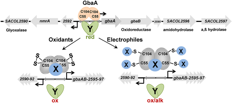
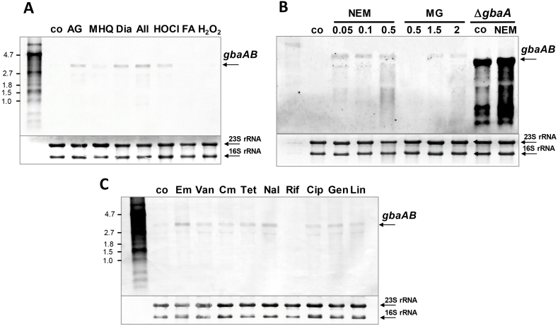
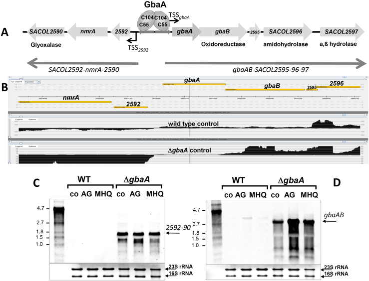
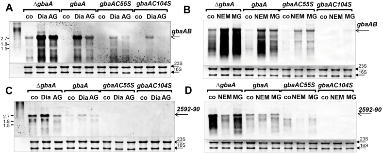
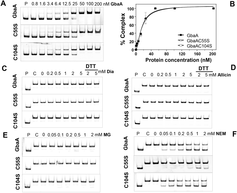
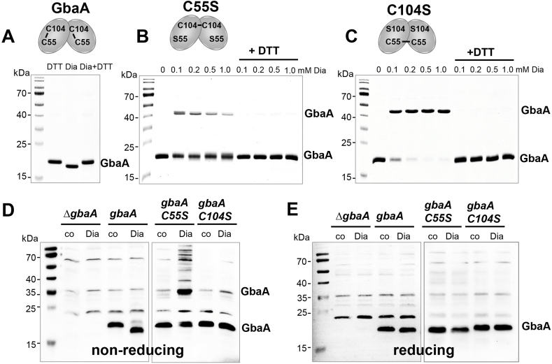
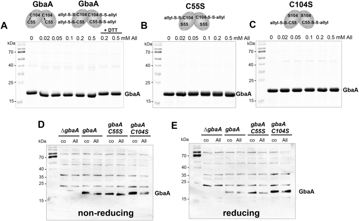
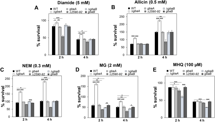
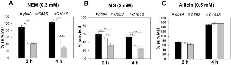
Staphylococcus aureus has to cope with oxidative and electrophile stress during host-pathogen interactions. The TetR-family repressor GbaA was shown to sense electrophiles, such as N-ethylmaleimide (NEM) via monothiol mechanisms of the two conserved Cys55 or Cys104 residues in vitro. In this study, we further investigated the regulation and function of the GbaA repressor and its Cys residues in S. aureus COL. The GbaA-controlled gbaAB-SACOL2595-97 and SACOL2592-nmrA-2590 operons were shown to respond only weakly 3-10-fold to oxidants, electrophiles or antibiotics in S. aureus COL, but are 57-734-fold derepressed in the gbaA deletion mutant, indicating that the physiological inducer is still unknown. Moreover, the gbaA mutant remained responsive to disulfide and electrophile stress, pointing to additional redox control mechanisms of both operons. Thiol-stress induction of the GbaA regulon was strongly diminished in both single Cys mutants, supporting that both Cys residues are required for redox-sensing in vivo. While GbaA and the single Cys mutants are reversible oxidized under diamide and allicin stress, these thiol switches did not affect the DNA binding activity. The repressor activity of GbaA could be only partially inhibited with NEM in vitro. Survival assays revealed that the gbaA mutant confers resistance under diamide, allicin, NEM and methylglyoxal stress, which was mediated by the SACOL2592-90 operon encoding for a putative glyoxalase and oxidoreductase. Altogether, our results support that the GbaA repressor functions in the defense against oxidative and electrophile stress in S. aureus. GbaA represents a 2-Cys-type redox sensor, which requires another redox-sensing regulator and an unknown thiol-reactive ligand for full derepression of the GbaA regulon genes.

Keywords: Allicin; Diamide; Electrophiles; GbaA; Staphylococcus aureus; Thiol switches.


Conflict of interest statement

The authors declare that there is no conflict of interest.

Figures

Image 1
Graphical abstract
Fig. 1
Fig. 1
Transcription of the gbaAB-SACOL2595-97 operon is weakly up-regulated by oxidative, electrophile and antibiotic treatments in S. aureus COL. Northern blot analysis was carried out using RNA isolated from S. aureus COL WT before (co) and 30 min after exposure to different thiol-reactive compounds (A, B) or 60 min after antibiotic treatments (C). For stress experiments, cells were treated with 5 μg/ml AGXX® (AG), 50 μM methylhydroquinone (MHQ), 2 mM diamide (Dia), 300 μM allicin (All), 1 mM HOCl, 0.75 mM formaldehyde (FA) and 10 mM H2O2 (A) or to 0.05–0.5 mM N-ethylmaleimide (NEM), 0.5–2 mM methylglyoxal (MG) for 30 min (B). For comparison of the weak transcriptional induction of the gbaAB operon after 0.5 mM NEM and 2 mM MG stress in the WT, the ΔgbaA mutant was analyzed under control and 0.5 mM NEM stress showing full derepression of the gbaAB operon in the control (B). For antibiotics experiments, S. aureus WT was exposed to 0.25 μg/ml erythromycin (Em), 0.5 μg/ml vancomycin (Van), 4 μg/ml chloramphenicol (Cm), 5 μg/ml tetracycline (Tet), 128 μg/ml nalidixic acid (Nal), 0.1 μg/ml rifampicin (Rif), 32 μg/ml ciprofloxacin (Cip), 2 μg/ml gentamicin (Gen) and 2 μg/ml linezolid (Lin) (C). The arrows point toward the size of the gbaAB-SACOL2595-97 specific operon transcript. The methylene blue stain is the RNA loading control indicating the 16S and 23S rRNAs. Band intensities of the gbaAB operon transcripts were quantified using ImageJ and the data shown in Figs. S4A–D.
Fig. 2
Fig. 2
Deletion of gbaA results in derepression of transcription of the downstream gbaAB-SACOL2595-97 and upstream SACOL2592-90 operons. (A, B) Transcriptional organization of the divergent gbaAB-SACOL2595-97 and SACOL2592-90 operons in S. aureus. The upstream SACOL2592-nmrA-2590 operon encodes for a putative glyoxalase and NAD+-dependent epimerase/dehydratase (NmrA). The downstream gbaAB operon encodes for the GbaA repressor, a putative short chain oxidoreductase, an amidohydrolase and an a,ß hydrolase. (B) Both operons are negatively regulated by GbaA as displayed by the RNA-seq data of S. aureus COL WT and the gbaA mutant under control conditions using Read-Explorer. (C, D) Transcription of the SACOL2592-90 (C) and gbaAB-SACOL2595-97 operons (D) was analyzed in S. aureus COL WT and gbaA mutant strains before (co) and 30 min after treatment with 5 μg/ml AGXX® (AG) and 50 μM MHQ using Northern blots. Both operons remained inducible by AGXX® and MHQ stress in the gbaA mutant. The arrows point toward the transcript sizes of the gbaAB and SACOL2592-90 operons. The methylene blue bands denote the 16S and 23S rRNAs as RNA loading controls below the Northern blots. Band intensities of the gbaAB and SACOL2592-90 operon transcripts were quantified using ImageJ and the data are shown in Figs. S5A and B. (For interpretation of the references to color in this figure legend, the reader is referred to the Web version of this article.)
Fig. 3
Fig. 3
Mapping of the 5′ ends of the gbaAB-SACOL2595-97 and SACOL2592-90 operons and the 9-6-9 bp inverted repeat as GbaA operator in S. aureus (A). 5′ RNA-seq was used to map TSS-1 and TSS-2 upstream of the divergent gbaAB and SACOL2592-90 operons, respectively, which is displayed with Read-Explorer. (B) The promoter sequence of the gbaAB operon and the 9-6-9 bp inverted repeat are highly conserved across different Staphylococcus species. The promoter regions were aligned using Clustal Omega and presented in Jalview. Intensity of the blue color gradient is based on 50% nucleotide sequence identity. (C) The conservation of the gbaA −10 promoter region and the GbaA operator is further displayed with WebLogo. (For interpretation of the references to color in this figure legend, the reader is referred to the Web version of this article.)
Fig. 4
Fig. 4
Northern blot analysis of transcription of the gbaAB-SACOL2595-97 and SACOL2592-90 operons under diamide, AGXX®, NEM and MG stress in the S. aureus COL gbaA mutant and the gbaA, gbaAC55S and gbaAC104S complemented strains. Transcription of the gbaAB-SACOL2595-97 (A,B) and SACOL2592-90 operons (C,D) was analyzed in the S. aureus gbaA deletion mutant and in the gbaA, gbaAC55S, gbaAC104S complemented strains before (co) and 30 min after treatment with 2 mM diamide (Dia), 5 μg/ml AGXX® (AG), 0.3 mM NEM and 2 mM MG using Northern blots. The arrows point toward the transcript sizes of the gbaAB-SACOL2595-97 or SACOL2592-90 operons. The methylene blue bands denote the 16S and 23S rRNAs as RNA loading controls below the Northern blots. Band intensities of the gbaAB and SACOL2592-90 operon transcripts were quantified using ImageJ and the data are shown in Figs. S6A–D.
Fig. 5
Fig. 5
The DNA binding activity of GbaA and the Cys mutant proteins is not inhibited under disulfide stress (diamide, allicin) and MG, but partially affected by NEM in vitro. (A) EMSAs were used to analyze the DNA binding activity of increasing concentrations of GbaA, GbaAC55S and GbaAC104S proteins to the 150 bp gbaA promoter probe. (B) The percentage of the GbaA-DNA complex formation was determined according to the band intensities of five biological replicates of the EMSAs in A) and quantified using Image J 1.48v. Dissociation constants (KD) were calculated as 15.24 nM, 15.24 nM and 15.79 nM for GbaA, GbaAC55S and GbaAC104S mutant proteins, respectively using the Graph prism software version 6.01. (C-F) The DNA binding activity of GbaA, GbaAC55S and GbaAC104S proteins was not affected by diamide, allicin and MG (C–E), but partially inhibited with NEM (F).
Fig. 6
Fig. 6
GbaA and the GbaA Cys mutants are oxidized to intra- and intermolecular disulfides by diamide in vitro and in vivo, respectively. (A–C) Purified GbaA was treated with 1 mM diamide (A), while the GbaAC55S (B) and GbaAC104S mutant proteins (C) were exposed to increasing concentrations of diamide for 15 min, followed by alkylation with 50 mM IAM for 30 min in the dark and separation by non-reducing SDS-PAGE. The non-reducing SDS-PAGE gels are stained with Coomassie Blue. To assess the reversibility, diamide-treated samples were reduced with 20 mM DTT for 15 min before alkylation and analysis by non-reducing SDS-PAGE. GbaA is oxidized to intramolecular C55-C104 disulfides by diamide as confirmed by MALDI-TOF MS (Fig. S7), while the C55S and C104S mutants form intermolecular disulfides as shown in the schematics above the gel images. (D, E) The S. aureus gbaA mutant and the gbaA complemented strain were treated with 5 mM diamide and the gbaAC55S and gbaAC104S complemented strains were exposed to 2 mM diamide for 30 min, alkylated with NEM and the protein extracts analyzed for thiol-oxidation of GbaA in vivo by non-reducing (D) and reducing (E) Western blot analysis with monoclonal anti-His6 tag antibodies. The protein loading controls are shown in Fig. S8.
Fig. 7
Fig. 7
GbaA is oxidized to intramolecular disulfides and S-thioallylations by allicin in vitro. (A–C) Purified GbaA (A), GbaAC55S (B) and GbaAC104S mutant proteins (C) were treated with increasing concentrations of allicin for 15 min, followed by alkylation with 50 mM IAM for 30 min in the dark and separation by non-reducing SDS-PAGE. The non-reducing SDS-PAGE gels are stained with Coomassie Blue. For the analysis of reversibility, allicin-treated samples were reduced by 20 mM DTT for 15 min, alkylated and subjected to non-reducing SDS-PAGE. GbaA was oxidized to intramolecular disulfides and S-thioallylations. The GbaA Cys mutants are S-thioallylated under allicin stress as revealed by MALDI-TOF MS (Fig. S7) and shown in the schematics above the images. (D, E) The S. aureus gbaA mutant and gbaA, gbaAC55S and gbaAC104S complemented strains were treated with 0.3 mM allicin stress for 30 min, alkylated with NEM and the protein extracts were used to analyze thiol-oxidation of GbaA in vivo by non-reducing (D) and reducing (E) Western blot analysis with monoclonal anti-His6 tag antibodies. The protein loading controls are shown in Fig. S8.
Fig. 8
Fig. 8
The GbaA regulon confers resistance under disulfide stress (diamide, allicin) and electrophiles (NEM, MG) in S. aureus. For survival assays, S. aureus COL WT, the gbaA, gbaB and SACOL2592-90 deletion mutants and gbaA, gbaB complemented strains were grown in RPMI medium until an OD500 of 0.5 and treated with 5 mM diamide (A), 0.5 mM allicin (B), 0.3 mM NEM (C), 2 mM MG (D) and 100 μM MHQ (E). CFUs were counted after plating 100 μl of serial dilutions onto LB agar plates after 2 and 4 h of stress exposure. The survival of treated cells was normalized to the untreated control, which was set to 100%. The results are from four biological replicates. Error bars indicate the standard deviation (SD) and the statistics was calculated using a Student's unpaired two-tailed t-test. Symbols are: *p ≤ 0.05, **p ≤ 0.01 and ***p ≤ 0.001.
Fig. 9
Fig. 9
The Cys55 and Cys104 residues of GbaA are required for NEM and MG tolerance. Survival assays were performed for the S. aureus gbaA mutant complemented with gbaA, gbaAC55S and gbaAC104S alleles. Strains were grown in RPMI until an OD500 of 0.5 and treated with 0.3 mM NEM (A), 2 mM MG (B) and 0.5 mM allicin (C) to determine CFUs after 2 and 4 h of stress exposure. The percentage survival was normalized to the control. Error bars represent the standard deviation (SD) calculated from three biological replicates. The statistics was determined using a Student's unpaired two-tailed t-test. Symbols are: *p ≤ 0.05, **p ≤ 0.01 and ***p ≤ 0.001.

Similar articles

Cited by

References

    1. Foster T.J. The Staphylococcus aureus "superbug. J. Clin. Invest. 2004;114(12):1693–1696. - PMC - PubMed
    1. Lowy F.D. Staphylococcus aureus infections. N. Engl. J. Med. 1998;339(8):520–532. - PubMed
    1. Boucher H.W., Corey G.R. Epidemiology of methicillin-resistant Staphylococcus aureus. Clin. Infect. Dis. 2008;46(Suppl 5):S344–S349. - PubMed
    1. Archer G.L. Staphylococcus aureus: a well-armed pathogen. Clin. Infect. Dis. 1998;26(5):1179–1181. - PubMed
    1. Chambers H.F., Deleo F.R. Waves of resistance: Staphylococcus aureus in the antibiotic era. Nat. Rev. Microbiol. 2009;7(9):629–641. - PMC - PubMed

Publication types