Review
. 2021 Nov:73:103642.
doi: 10.1016/j.ebiom.2021.103642. Epub 2021 Oct 19.

Off balance: Interferons in COVID-19 lung infections

Affiliations
Review

Off balance: Interferons in COVID-19 lung infections

Milena Apetito Akamatsu et al. EBioMedicine. 2021 Nov.

Abstract

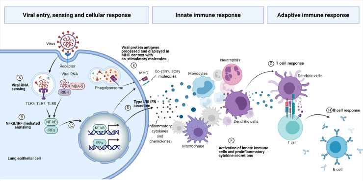
Interferons are innate and adaptive cytokines involved in many biological responses, in particular, viral infections. With the final response the result of the balance of the different types of Interferons. Cytokine storms are physiological reactions observed in humans and animals in which the innate immune system causes an uncontrolled and excessive release of pro-inflammatory signaling molecules. The excessive and prolonged presence of these cytokines can cause tissue damage, multisystem organ failure and death. The role of Interferons in virus clearance, tissue damage and cytokine storms are discussed, in view of COVID-19 caused by Severe Acute Respiratory Syndrome Coronavirus 2 (SARS-CoV-2). The imbalance of Type I, Type II and Type III Interferons during a viral infection contribute to the clinical outcome, possibly together with other cytokines, in particular, TNFα, with clear implications for clinical interventions to restore their correct balance.

Keywords: COVID-19; Cytokine Storms; Interferons; SARS-CoV-2; TNFα.


Conflict of interest statement

Declaration of Competing Interest The authors declare that the research was conducted in the absence of any commercial or financial relationships that could be construed as a potential conflict of interest.

Figures

Figure 1
Figure 1
– Type I, II and III Interferons, their receptors and signalings. (A) The Type I, II and III IFN ligands and their receptors; (B) Kinases associated with the Type I, II and III receptors that mediate IFN signallings through receptor activation by ligand binding. Jak1 (Janus Kinase 1) interacts with STAT1 (Signal Transducer and Activator of Transcription 1) as well as to IFNRA1 (Interferon receptor alpha 1). Tyk2 (Tyrosine kinase 2) interacts to STAT2 as well as to IFNRA2. IFNRA1 forms a heterodimer with IFNRA2 that binds with Type I IFN; Jak1 and STAT1 also interacts to IFNLR1. IL-10RB (Interleukin-10 receptor B) also interacts with Tyk2 and STAT2. IFNLR1 forms a heterodimer with IL-10RB that binds to Type III IFNs. Jak1 and STAT1 also interacts to IFNGR1 and JAK2 and STAT1 also interacts with IFNGR2. IFNGR1 forms a heterodimer with IFNGR2 that binds to Type II IFNs; (C) Upon ligand binding and activation, the STAT1 or STAT2 are phosphorylated and the STAT1 homodimers go to the nucleous and activate the transcription of genes with the GAS (gama interferon activation sites) promoters; STAT1 and STAT2 heterodimers binds to IRF9 (interferon regulatory factor 9) and the complex goes to the nucleous and activates the the transcription of genes with the ISRE (Interferon-stimulated responsive element) promoters. Some commom ISGs (trafd1, TNF receptor-associated factor -type zinc finger domain containing 1; parp 9, Poly(ADP-ribose) polymerase family member 9; parp12, Poly(ADP-ribose) polymerase family member 12; parp14, Poly(ADP-ribose) polymerase family member 14; nmi, N-myc and STAT interactor; epsti1, epithelial stromal interaction 1; eif2ak2, eukaryotic translation initiation factor 2 alpha kinase 2; irf1, interferon regulatory factor 1; irf7, interferon regulatory factor 7; irf8, interferon regulatory factor 8; tmem67, transmembrane protein 67; tmem140, transmembrane protein 140; tmem173, transmembrane protein 173; dtx3l, deltex E3 ubiquitin-protein ligase; samhd1, SAM (sterile alpha-motif) and HD (histidine-aspartate) domain containing deoxynucleoside triphosphate truphosphohydrolase 1; stat1, signal transducer and activator of transcription 1; cd274, cluster of differentiation 274) induced by Type I, II and III IFNs are indicated by * [69,70]. (Figure created with BioRender.com).
Figure 2
Figure 2
– Schematic view of the viral entry, sensing and host immune response. The RNA virus bind to the cellular receptor and is internalized. Pattern Recognition Receptors (PRRs) sense the viral RNA (A) and activate cellular signaling mediated by NF-κB and IRFs transcription factors (B) to the nucleous (C), activating expression of specific genes, comprising Type I and Type III IFNs and proinflammatory cytokines (D). Viral protein antigens are also processed and displayed in MHC context with co-stimulatory molecules (E). The proinflammatory cytokines and chemokines are responsible for the cellular innate response, with pronounced infiltration of monocytes and neutrophils (F). Activated dendritic cells present the antigens to the adaptive T cells (G) that will secrete cytokines to mature B cells to produce and secrete immunoglobulins (H) (Figure created by BioRender.com).
Figure 3
Figure 3
– Respiratory tract infection by SARS-CoV-2. Infection occurs at the epithelial cells of the respiratory tract and the infection may be inhibited by pre-existing cross-reactive antibodies resulted from previous infections with seasonal viruses (A). The infection causes inflammation in the respiratory tract with the secretion of proinflammatory cytokines (B), activation of the endothelial cells (C) and expression of NK receptor ligands (MICA/B, MHC class I chain related protein A or B) on respiratory epithilium (C´). The activated endothelium promotes the infiltration of NK-like T cells expressing NK (Natural Killer) receptors (NKR) exemplified by NKG2D on the cell surface from the capillaries (D) to the respiratory tract epithelium. The infiltrating NK-like T cells binds to NKR receptor ligands (MICA/B) and induce TCR-independent killing of epithelium cells expressing the NKR ligands (E). In response to viral infections, epithelium cells secrete Type I or Type III IFNs. In severe cases of the diseases, the presence of autoantibodies against IFNα and IFNω (F) was observed and associated with higher morbidity, resulting in more viral infections. It was also observed an increase in the secretion of Type III IFN (IFNλ)(F). IFNλ impairs lung epithelial cell proliferation and tissue repair mediated by the expression of the tumor suppressor p53 gene and protein pathway. Cell death programs (PANoptosis) induced by cytokine storms, in special by IFNγ and TNFα, perpetuates the local cytokine storms killing more epithelial cells in the respiratory tract (G) and the cytokine storms propagate (H) to other organs and tissues, provoking cytokine shock syndromes. Acute respiratory distress syndrome (ARDS) will be observed in the patient due to lung damage as well as multi-organ failures (I) due to systemic spread of the proinflammatory cytokines, in special of IFNγ and TNFα. The cytokine shock syndromes can be identified by clinical markers as listed in the figure (J). Susceptibility to bacterial superinfections is increased in damaged respiratory tract (K) due to cell killings by NK-like T cells (E) and by PANoptosis (G) in concert with inhibition of epithelial cell proliferation and repair by IFNλ (F). Cytokine shock sindrome markers: RBC, red blood cells; HCT, hematocrit; Hb, hemoglobin; PCT, Procalcitonin; LDH, lactate dehydrogenase; ALT, alanine aminotransferase; AST; aspartate aminotransferase; BUN, blood urea nitrogen. (Figure created with BioRender.com).

References

    1. Walter MR. The Role of Structure in the. Biology of Interferon Signaling. Front. Immunol. 2020;11 doi: 10.3389/fimmu.2020.606489. - DOI - PMC - PubMed
    1. Schoggins JW. Interferon-Stimulated Genes: What Do They All Do? Annu. Rev. Virol. 2019;6(1):567–584. doi: 10.1146/annurev-virology-092818-015756. - DOI - PubMed
    1. Onomoto K, Onoguchi K, Yoneyama M. Regulation of RIG-I-like receptor-mediated signaling: interaction between host and viral factors. Cell. Mol. Immunol. 2021;18:1–17. doi: 10.1038/s41423-020-00602-7. - DOI - PMC - PubMed
    1. Au-Yeung N, Horvath CM. Transcriptional and chromatin regulation in interferon and innate antiviral gene expression. Cytokine Growth Factor Rev. 2018;44:11–17. - PMC - PubMed
    1. Monchatre-Leroy E., Boué F., Boucher J.-M., Renault C., Moutou F., Ar Gouilh M., Umhang G. Identification of Alpha and Beta Coronavirus in Wildlife Species in France: Bats, Rodents, Rabbits, and Hedgehogs. Viruses. 2017;9:364. doi: 10.3390/v9120364. - DOI - PMC - PubMed

MeSH terms