

Evaluation of acupuncture treatments of postpartum female pelvic floor dysfunction by four-dimensional transperineal pelvic floor ultrasound
- PMID: 34678860
- PMCID: PMC8542121
- DOI: 10.1097/MD.0000000000027236
Evaluation of acupuncture treatments of postpartum female pelvic floor dysfunction by four-dimensional transperineal pelvic floor ultrasound
Abstract
Introduction: In the present investigation, a systematic evaluation of the clinical treatment performance of diagnosed with pelvic floor dysfunction is explored. By comparing the 4Dtransperineal pelvic floor ultrasound images with the acupuncture treatment performance of the patients, an evaluation system with various parameters is established to provide critical information to guide the clinical treatment fpostpartum female pelvic floor dysfunction (FPFD).
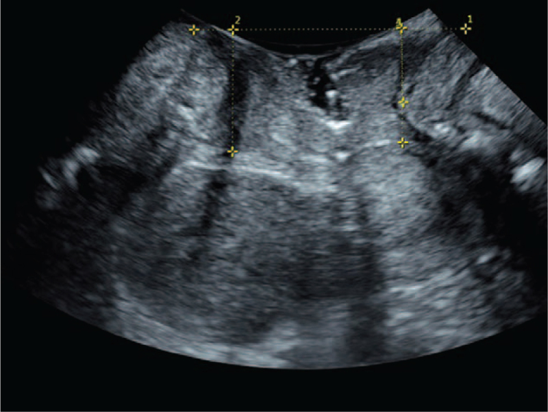
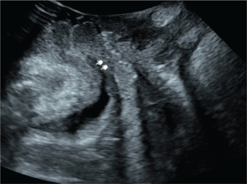
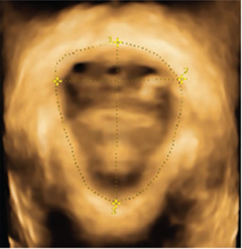
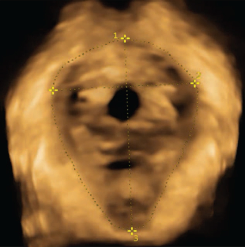
Methods: Eighty patients diagnosed with FPFD are divided into 2 groups. After the designated treatment to the patients, they are carefully examined using transperineal pelvic floor ultrasound. The shape and activity of bladder neck, cervix and rectum anal canal under resting, anal sphincter and Valsalva movements are observed and recorded. The morphology and continuous shape of levator ani muscle in different states after 4D image reconstruction are obtained.
Results: After the acupuncture treatment, the bladder neck descent is decreased by 3.8 cm and the anal levator muscle area is decreased by 3.4 cm2 comparing with the control group. The anal levator muscle hole diameter is decreased by 0.3 cm, while the anterior and posterior diameter is reduced by 0.5 cm. Reduced possibility of cystocele and uterine prolapse is demonstrated by X2 test. These changes upon acupuncture therapy are in line with the improved conditions of the patients, indicating these parameters can help evaluate the therapy performance.
Conclusion: 4D pelvic floor ultrasound imaging provides objective and quantified information for the clinical diagnosis and treatment of FPFD and the assessment of therapy efficacy, making it a promising novel method in practical applications.
Copyright © 2021 the Author(s). Published by Wolters Kluwer Health, Inc.
Conflict of interest statement
The authors have no conflicts of interests to disclose.
Figures
Similar articles
-
Clinical value of transperineal ultrasound in evaluating the effects of different delivery methods on the primipara pelvic floor structure and function.Sci Rep. 2024 Oct 14;14(1):23980. doi: 10.1038/s41598-024-75014-y. Sci Rep. 2024. PMID: 39402151 Free PMC article.
-
Assessment of levator hiatal area using 3D/4D transperineal ultrasound in women with deep infiltrating endometriosis and superficial dyspareunia treated with pelvic floor muscle physiotherapy: randomized controlled trial.Ultrasound Obstet Gynecol. 2021 May;57(5):726-732. doi: 10.1002/uog.23590. Ultrasound Obstet Gynecol. 2021. PMID: 33428320 Clinical Trial.
-
Ultrasound Diagnosis of Levator Ani Hiatus Enlargement and Cystocele in Standing and Supine Positions in the Postpartum Period.J Ultrasound Med. 2025 Apr;44(4):681-689. doi: 10.1002/jum.16627. Epub 2024 Dec 5. J Ultrasound Med. 2025. PMID: 39635975 Free PMC article.
-
[Postpartum levator ani muscle injuries. Diagnosis and treatment].Ginekol Pol. 2015 Jan;86(1):67-71. doi: 10.17772/gp/1902. Ginekol Pol. 2015. PMID: 25775878 Review. Polish.
-
Levator ani muscle avulsion in patients with pelvic floor dysfunction - Does it help in understanding pelvic organ prolapse?Eur J Obstet Gynecol Reprod Biol. 2022 Dec;279:140-145. doi: 10.1016/j.ejogrb.2022.09.033. Epub 2022 Sep 30. Eur J Obstet Gynecol Reprod Biol. 2022. PMID: 36343586 Review.
Cited by
-
Transperineal pelvic floor ultrasound for assessing posterior pelvic injury and prolapse in postpartum women.Am J Transl Res. 2023 Oct 15;15(10):6170-6179. eCollection 2023. Am J Transl Res. 2023. PMID: 37969208 Free PMC article.
-
Effect of 3D Animation Combined with Teach-Back Health Education on Pelvic Floor Muscle Training in LARS Patients: A Randomized Controlled Trial.J Nurs Manag. 2023 Feb 24;2023:6847933. doi: 10.1155/2023/6847933. eCollection 2023. J Nurs Manag. 2023. PMID: 40225656 Free PMC article. Clinical Trial.
-
Image Enhancement Algorithm-Based Ultrasound on Pelvic Floor Rehabilitation Training in Preventing Postpartum Female Pelvic Floor Dysfunction.Comput Math Methods Med. 2022 Apr 19;2022:8002055. doi: 10.1155/2022/8002055. eCollection 2022. Comput Math Methods Med. 2022. PMID: 35495879 Free PMC article. Clinical Trial.
-
Efficacy of Acupuncture in Post-partum With Diastasis Recti Abdominis: A Randomized Controlled Clinical Trial Study Protocol.Front Public Health. 2021 Dec 13;9:722572. doi: 10.3389/fpubh.2021.722572. eCollection 2021. Front Public Health. 2021. PMID: 34966711 Free PMC article.
-
Efficacy of Warm Acupuncture Therapy Combined with Kegel Exercise on Postpartum Pelvic Floor Dysfunction in Women.Int Urogynecol J. 2024 Mar;35(3):599-608. doi: 10.1007/s00192-023-05698-9. Epub 2024 Jan 18. Int Urogynecol J. 2024. PMID: 38236284 Free PMC article. Clinical Trial.
References
-
- Hoyte L, Schierlitz L, Zou K, Flesh G, Fielding JR. Two- and 3-dimensional MRI comparison of levator ani structure, volume, and integrity in women with stress incontinence and prolapse. Am J Obstet Gynecol 2001;185:11–9. - PubMed
-
- Alt CD, Hampel F, Radtke JP, et al. . Early postpartum pelvic floor changes in primiparous women after vaginal delivery using 3T MRI. Neurourol Urodynamics 2017;36:2064–73.
-
- Santoro GA. Imaging the pelvic floor. Tech Coloproctol 2017;21:497–9. - PubMed
-
- Weinstein MM, Jung SA, Pretorius DH, Nager CW, den Boer DJ, Mittal RK. The reliability of puborectalis muscle measurements with 3-dimensional ultrasound imaging. Am J Obstet Gynecol 2007;197:68 e61–6. - PubMed
Publication types
MeSH terms
LinkOut - more resources
Full Text Sources
Medical
