. 2021 Sep 25;9(10):237.
doi: 10.3390/toxics9100237.

The Peculiar Estrogenicity of Diethyl Phthalate: Modulation of Estrogen Receptor α Activities in the Proliferation of Breast Cancer Cells

Affiliations

The Peculiar Estrogenicity of Diethyl Phthalate: Modulation of Estrogen Receptor α Activities in the Proliferation of Breast Cancer Cells

Marco Fiocchetti et al. Toxics. .

Abstract

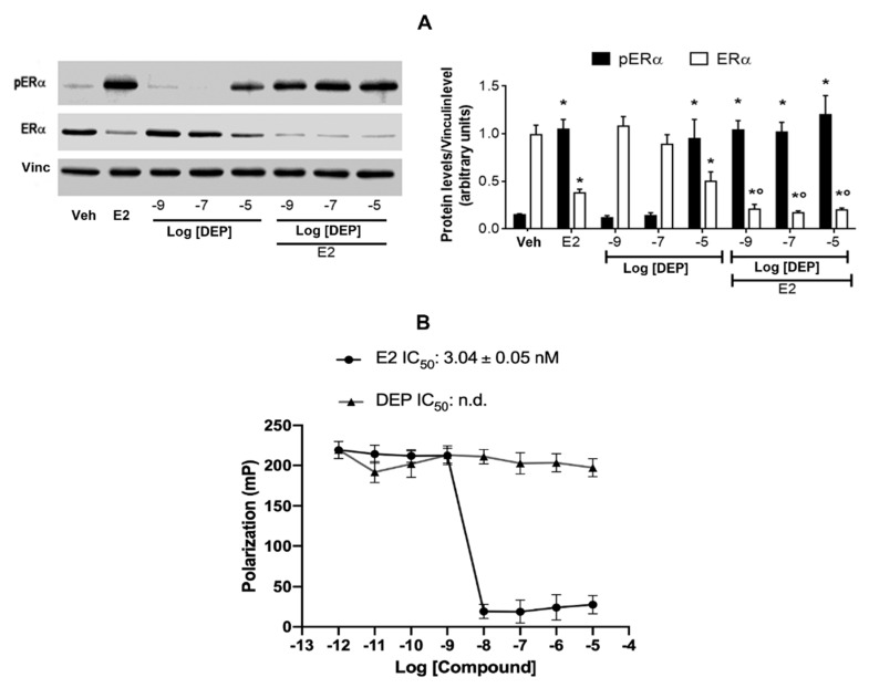
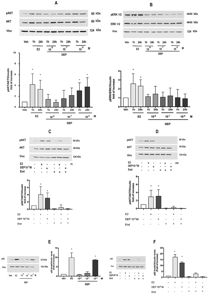
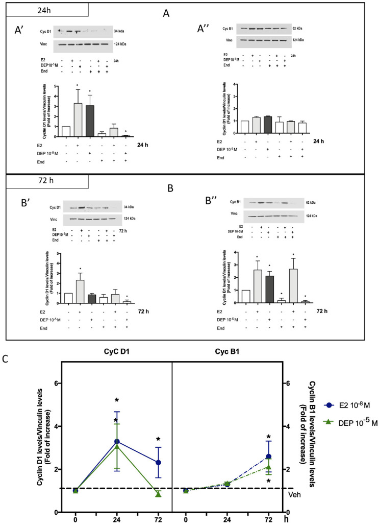
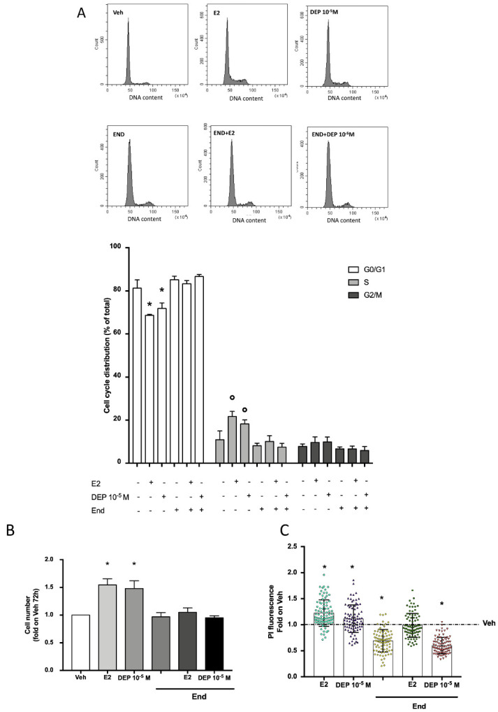
Phthalates comprise a group of synthetic chemicals present in the environment because of their wide use as plasticizers and as additives in products for personal care. Among others, diethyl phthalate (DEP) is largely used in products for infants, children, and adults, in which its exposure has been correlated with an increased risk of breast cancer. The adverse health outcomes deriving from phthalate exposure have been associated with their activity as endocrine disruptors (EDCs) of the steroid and thyroid hormone signaling by affecting developmental and reproductive health, and even carcinogenicity. However, the estrogen disruptor activities of DEP are still controversial, and the mechanism at the root of the estrogenic-disrupting action of DEP remains to be clarified. Here, we evaluated the DEP mechanism of action on the activation status of estrogen receptor α (ERα) by analyzing the receptor's phosphorylation as well as both nuclear and extra-nuclear pathways triggered by the receptor to modulate the proliferation of breast cancer cells. Although DEP does not bind to ERα, our results suggest that this phthalate ester exerts multiple parallel interactions with ERα signaling and emphasize the importance to determine an appropriate battery of in vitro methods that will include specific molecular mechanisms involved in the endocrine disruption.

Keywords: breast cancer cell proliferation; diethyl phthalate; endocrine disruptors; estrogen receptor α; signal transduction pathways.


Conflict of interest statement

The authors declare no conflict of interest.

Figures

Figure 1
Figure 1
ERα activation and in vitro binding assays. Western blot (left panel) and corresponding densitometric analysis (right panel) of Ser118-phosphorylated ERα and total ERα protein level in MCF-7 treated for 1 h with the endogenous ERα ligand E2 (10−8 M) as positive control, and a dose curve of DEP at 10−9, 10−7, and 10−5 M in the presence or absence of E2 co-treatment (A). The vinculin levels were used as an internal control of protein loading. Data are means ± SD of at least four experiments. p < 0.01 was determined by the ANOVA test followed by the Bonferroni post-test vs. Veh (*) and E2 (°). In vitro ERα competitive binding assay for the endogenous ligand E2 and DEP performed over the 10−12–10−5 M range by using fluorescent E2 as a tracer. Inhibitor concentration (IC50- nM) is indicated in the panel for each compound (B).
Figure 2
Figure 2
DEP activates nuclear and extra-nuclear rapid ERα signals. DEP dose-dependent effect on phosphorylation of AKT (A) and ERK1/2 (B) analyzed at short (1 h) and long (24 h) time exposures. Data show the Western blot image representative of at least four different experiments (upper panel) and the densitometric analysis (bottom panel). Western blot analysis of pAKT levels (upper panel) and densitometric analysis (bottom panel) in MCF-7 treated with DEP 10−5 M for 1 h (C) or 24 h (D) with or without cell pre-treatment with specific ERα inhibitor endoxifen (End; 10−6 M, 1 h before). Western blot (left panel) and corresponding densitometric analysis (right panel) of the E2-responsive protein pS2 in MCF-7 cells treated for 24 h with a dose curve of DEP (10−9, 10−7, 10−5 M) (E) or with DEP 10−5 M in the presence or absence of End (10−6 M, 1 h pre-treatment) (F). E2 in the presence and/or absence of End pre-treatment was used as internal positive control throughout all the experiments. The amount of protein was normalized in comparison with vinculin levels or with total AKT or total ERK1/2 and vinculin levels. Data are means ± SD of at least four experiments. p < 0.01 was determined by the ANOVA test followed by the Bonferroni post-test vs. Veh (-,-,-) (*).
Figure 3
Figure 3
ERα-dependent effect of DEP on cyclin expression. MCF-7 cells were treated with DEP 10−5 M for 24 h (A) or 72 h (B) in the presence or absence of End (10−6 M, 1 h before) pre-stimulation, and the protein expression levels of cyclin D1 (Cyc D1; A’,B’) and cyclin B1 (Cyc B1; A”,B”) were evaluated by Western blot. Data show representative Western blot images of at least four experiments (upper panels) and the corresponding densitometric analysis (bottom panels). E2 with or without End pre-treatment was used as positive control. Analysis of expression of cyclin D1 (left box) and cyclin B1 (right box) as a function of the time of DEP 10−5 M exposure (C). The level of vinculin was used for protein normalization. Data are means ± SD of at least four experiments. p < 0.01 was determined by the ANOVA test followed by the Bonferroni post-test vs. Veh (-,-,-) (*).
Figure 4
Figure 4
The effect of DEP on ERα-dependent DNA synthesis, cell cycle progression, and proliferation. Cell cycle analysis in MCF-7 cells treated with DEP 10−5 M for 24 h with or without pre-stimulation with End (10−6 M, 1 h before). Upper panels show flow cytometry analysis for cell cycle distribution of MCF-7 cells under different treatment conditions. The bottom panel shows the percentage of cells in G0/G1, S, and G2/M phases with respect to different stimulations. Histograms are representative of at least three independent experiments (A). Viable cell number (B) and analysis of total DNA content obtained from propidium iodide (PI) assay (B) of MCF-7 cells treated with DEP 10−5 M for 72 h in the presence or absence of End pre-treatment (10−6 M, 1 h before). Data are shown as fold of increase in Vehicle condition represented by the blank column (B) or as the dotted line (C). E2 stimulation with or without End pre-stimulation was used as positive control. p < 0.01 was determined by the ANOVA test followed by the Bonferroni post-test vs. G0/G1 phase Veh (-,-,-) and Veh (B,C) (*) or vs. S phase Veh (-,-,-) (°).

References

    1. Ascenzi P., Bocedi A., Marino M. Structure–function relationship of estrogen receptor α and β: Impact on human health. Mol. Asp. Med. 2006;27:299–402. doi: 10.1016/j.mam.2006.07.001. - DOI - PubMed
    1. Cooke P.S., Nanjappa M.K., Ko C., Prins G.S., Hess R. Estrogens in Male Physiology. Physiol. Rev. 2017;97:995–1043. doi: 10.1152/physrev.00018.2016. - DOI - PMC - PubMed
    1. Bulzomi P. Environmental endocrine disruptors: Does a sex-related susceptibility exist? Front. Biosci. 2011;16:2478–2498. doi: 10.2741/3867. - DOI - PubMed
    1. Guercio G., Saraco N., Costanzo M., Marino R., Ramirez P., Berensztein E., Rivarola M.A., Belgorosky A. Estrogens in Human Male Gonadotropin Secretion and Testicular Physiology From Infancy to Late Puberty. Front. Endocrinol. 2020;11:72. doi: 10.3389/fendo.2020.00072. - DOI - PMC - PubMed
    1. Marino M., Pellegrini M., La Rosa P., Acconcia F. Susceptibility of estrogen receptor rapid responses to xenoestrogens: Physiological outcomes. Steroids. 2012;77:910–917. doi: 10.1016/j.steroids.2012.02.019. - DOI - PubMed

LinkOut - more resources