Review
. 2021 Sep 27;11(10):1279.
doi: 10.3390/brainsci11101279.

Cortical Visual Impairment in Childhood: 'Blindsight' and the Sprague Effect Revisited

Affiliations
Review

Cortical Visual Impairment in Childhood: 'Blindsight' and the Sprague Effect Revisited

Gerry Leisman et al. Brain Sci. .

Abstract

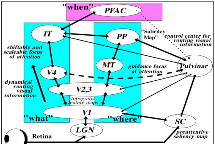
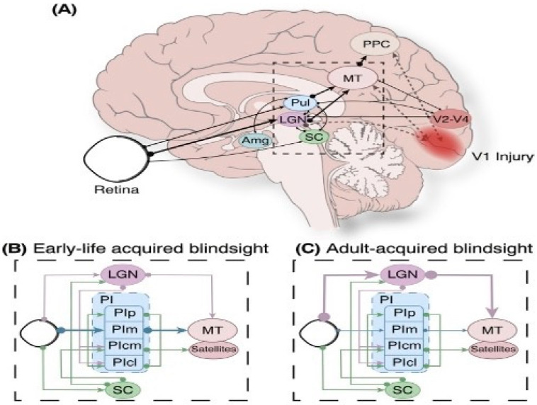
The paper discusses and provides support for diverse processes of brain plasticity in visual function after damage in infancy and childhood in comparison with injury that occurs in the adult brain. We provide support and description of neuroplastic mechanisms in childhood that do not seemingly exist in the same way in the adult brain. Examples include the ability to foster the development of thalamocortical connectivities that can circumvent the lesion and reach their cortical destination in the occipital cortex as the developing brain is more efficient in building new connections. Supporting this claim is the fact that in those with central visual field defects we can note that the extrastriatal visual connectivities are greater when a lesion occurs earlier in life as opposed to in the neurologically mature adult. The result is a significantly more optimized system of visual and spatial exploration within the 'blind' field of view. The discussion is provided within the context of "blindsight" and the "Sprague Effect".

Keywords: Sprague Effect; blindsight; cortical blindness; cortical visual impairment; infant vision; vision.


Conflict of interest statement

The authors declare no conflict of interest.

Figures

Figure 1
Figure 1
Diffusion tensor tractography of (A) optic radiations of left hemisphere evidencing a pathway avoiding an enlarged left ventricle projecting to the calcarine cortex. (B) bilateral optic radiations with the fibers of the left hemisphere proceeding laterally and anteriorly in contradistinction to the non-lesioned hemisphere. (C) Optic radiations of the right hemisphere appear to follow a normal trajectory (after Guzzetta et al. [56] with permission).
Figure 2
Figure 2
Major visual processing pathways of the primate brain considered in Gross et al.’s [99] model. Information from the retino-geniculostriatal pathway enters the visual cortex through area V1 and then proceeds through a hierarchy of visual areas that can be subdivided into two major functional pathways. The so-called “what”-pathway leads through V4 and the inferotemporal cortex (IT) and is mainly concerned with object-feature identification, regardless of position or size. V4 is the third area in the ventral stream obtaining strong feedforward signals from V2. Additionally, it receives projections directly from V1. The “where” pathway leads into the posterior parietal areas (PP) and is concerned with the locations and spatial relationships among objects, regardless of their identity. The “when” pathway involves the integration of signals from “What” and “Where” allowing for preplanning of movement and therefore response. (PFAC, prefrontal association cortex; IT, inferotemporal cortex; PP, posterior parietal areas; MT, middle temporal visual area; LGN, lateral geniculate nucleus; SC, superior colliculus) (after Gross et al. [99] with permission).
Figure 3
Figure 3
Potential mechanisms of neuroplasticity-based functional reorganization supporting normal visual function in congenitally brain-damaged individuals. (a) Represents damage to the PVC with functioning tissue existing within the lesion (b) aa reorganization occurring in regions external to the accepted boundaries of the PVC; (c) the geniculostriatal pathway bypassing the lesion and projecting to the calcarine cortex (after Guzzetta et al. [47] with permission).
Figure 4
Figure 4
Subtle deficits and residual functions in visual fields with CVI. The phenomenon of blindsight occurs when otherwise blind individuals can correctly guess that stimuli were presented without the individual’s awareness of them. CVI individuals can correctly respond to visual stimuli, but they report seeing nothing (unconsciously seeing without knowing). These are areas in the visual field which are neither blind nor seeing normally (areas that are blind (black), partially impaired (grey), or normal (white)). Visual fields typically have different “shades of grey” where function is neither completely lost nor normal. Here blind individuals only occasionally respond to stimulation. With repeated testing, these visual field regions are variably responsive and are associated with increased thresholds and longer response time. X and Y axes represent the degree of angular subtense from central fixation. The concealed deficits in the “seeing field” effectively render individuals “sightblind”, measured by examinations that can measure higher cognitive dysfunctions. Especially clinically relevant is the grey “area of residual vision”, where vision is neither normal nor absent. These are the regions of the visual field with the greatest recovery potential. (from Sabel et al. [146], with permission).
Figure 5
Figure 5
Subcortical pathways capable of facilitating blindsight. (A) Medial view demonstrating retinal extrastriatal region feedforward pathways in the posterior parietal cortex. Opaque pathways designate V1 injury neuronal degeneration. (B,C) Hypothesized subcortical pathways enabling blindsight after damage in V1 in the child’s (B) and in the adult brain (C). The strength of the projection is represented by line thickness. (Abbreviations: Amg, Amygdala; LGN, lateral geniculate nucleus; MT, middle temporal area; PI, inferior pulvinar; PIcl, caudolateral division of inferior pulvinar; PIcm, centromedial division of inferior pulvinar; PIm, medial division of inferior pulvinar; PIp, posterior division of inferior pulvinar; PPC, posterior parietal cortex; Pul, pulvinar; SC, superior colliculus) (from Fox at al., [149] with permission).

References

    1. Chang M.Y., Borchert M.S. Advances in the evaluation and management of cortical/cerebral visual impairment in children. Surv. Ophthalmol. 2020;65:708–724. doi: 10.1016/j.survophthal.2020.03.001. - DOI - PubMed
    1. Khan R.I., O’Keefe M., Kenny D., Nolan L. Changing pattern of childhood blindness. Ir. Med. J. 2007;100:458–461. - PubMed
    1. Kong L., Fry M., Al-Samarraie M., Gilbert C., Steinkuller P.G. An update on progress and the changing epidemiology of causes of childhood blindness worldwide. J. Am. Assoc. Pediatr. Ophthalmol. Strabismus. 2012;16:501–507. doi: 10.1016/j.jaapos.2012.09.004. - DOI - PubMed
    1. National Institutes of Health. [(accessed on 11 September 2021)];2021 Available online: https://www.nei.nih.gov/about/news-and-events/news/vision-loss-children-....
    1. Flanagan N.M., Jackson A.J., Hill A.E. Visual impairment in childhood: Insights from a community-based survey. Child Care Health Dev. 2003;29:493–499. doi: 10.1046/j.1365-2214.2003.00369.x. - DOI - PubMed

LinkOut - more resources