Review
. 2021 Sep 28;11(10):1789.
doi: 10.3390/diagnostics11101789.

The Role of Endoscopic Ultrasound in the Diagnosis of Gallbladder Lesions

Affiliations
Review

The Role of Endoscopic Ultrasound in the Diagnosis of Gallbladder Lesions

Senju Hashimoto et al. Diagnostics (Basel). .

Abstract

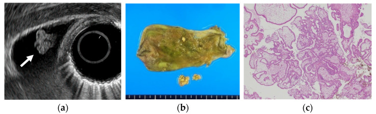
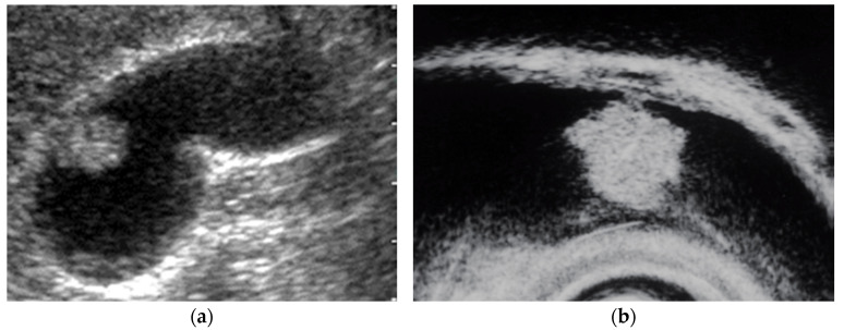
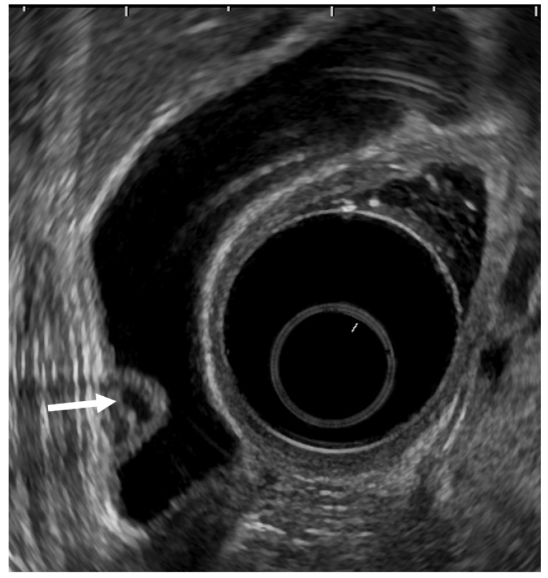
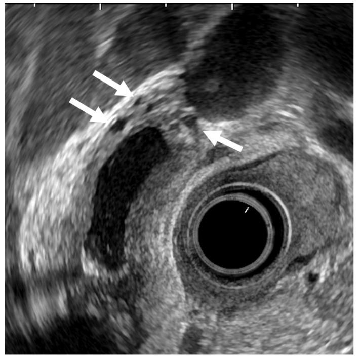
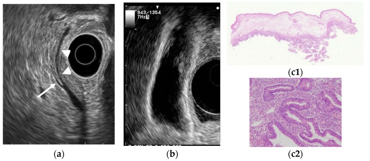
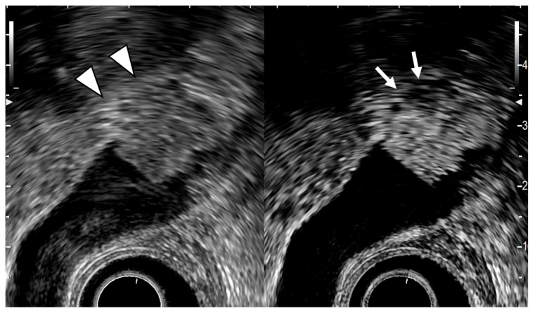
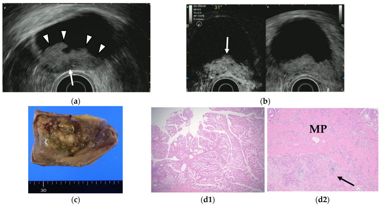
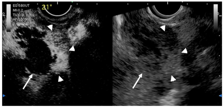
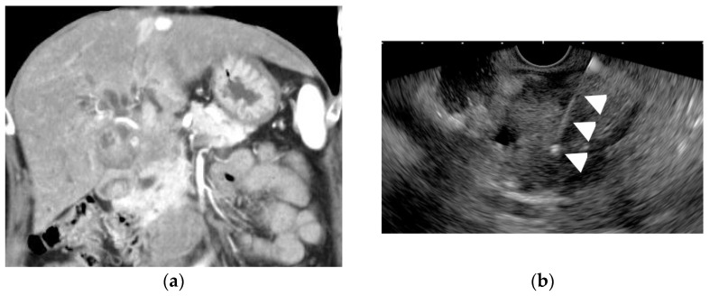
Gallbladder (GB) diseases represent various lesions including gallstones, cholesterol polyps, adenomyomatosis, and GB carcinoma. This review aims to summarize the role of endoscopic ultrasound (EUS) in the diagnosis of GB lesions. EUS provides high-resolution images that can improve the diagnosis of GB polypoid lesions, GB wall thickness, and GB carcinoma staging. Contrast-enhancing agents may be useful for the differential diagnosis of GB lesions, but the evidence of their effectiveness is still limited. Thus, further studies are required in this area to establish its usefulness. EUS combined with fine-needle aspiration has played an increasing role in providing a histological diagnosis of GB tumors in addition to GB wall thickness.

Keywords: EUS-guided fine-needle aspiration (EUS-FNA); contrast-enhanced EUS; differential diagnosis; endoscopic ultrasound (EUS); gallbladder carcinoma; polypoid lesion; staging of gallbladder carcinoma; wall-thickening.


Conflict of interest statement

The authors declare no conflict of interest.

Figures

Figure 1
Figure 1
Cholesterol polyp. (a) EUS shows a cholesterol polyp as a granular-surfaced pedunculated lesion. The internal echo is heterogeneous with a hyperechoic spot (arrow). (b) Photograph of the gross pathologic specimen after cholecystectomy shows a yellowish polyp. (c) H-E stain of the specimen demonstrates an aggregation of foamy cells under the epithelium.
Figure 2
Figure 2
Pedunculated GB carcinoma. (a) TUS image shows a relatively smooth surface, solid internal echogenicity polyp, but TUS does not depict the nature of the base of the lesion. (b) EUS image shows a pedunculated lesion. This lesion was GB adenocarcinoma with invasion depth pT1a (M) as a result of surgery.
Figure 3
Figure 3
EUS image ADM (localized type). EUS shows localized ADM as a sessile polypoid lesion with anechoic areas (arrow) corresponding to RAS proliferation. The surface is relatively smooth.
Figure 4
Figure 4
EUS image of ADM (diffuse type). The GB wall is diffusely thickened, and the layers of a thickened GB wall are preserved. Some anechoic areas (arrows) are visualized in the GB thickened wall.
Figure 5
Figure 5
GB carcinoma is associated with pancreaticobiliary maljunction without biliary dilatation. (a) EUS shows the bile duct (arrowhead) and main pancreatic duct (arrow) communicated inside the pancreas. (b) EUS shows the irregular GB wall-thickening on fundus without wall layer structure disruption. (c1,c2) H–E stain of the specimen demonstrates adenocarcinoma with tumor in situ stage.
Figure 6
Figure 6
EUS image of GB carcinoma. The conventional EUS (left) shows that a broad-based elevated lesion is found at the fundus of the GB with hypoechoic (arrowhead) in the deep part of the lesion and rupture of the lateral hyperechoic layer. In the contrast-enhanced EUS (right), the contrast effect of most of the lesion is good, but the deep part of the lesion is poorly contrasted (arrow). It can be diagnosed from these findings as GB carcinoma with invasion depth T3a (SE).
Figure 7
Figure 7
GB carcinoma. (a) Conventional EUS shows elevated lesions with conspicuous surface irregularities (arrowheads) observed in the gallbladder body. A hypoechoic region is observed in the deep part of the lesion (arrow), and the outer hyperechoic layer is also irregular, suggesting infiltration into the subserosal layer. (b) The contrast-enhanced harmonic EUS image after the injection of Sonazoid® shows that lesions in the gallbladder body (arrow) have a strong heterogeneous staining effect from the early stage of contrast enhancement. (Left contrast-enhanced harmonic mode, right B-mode) (c) Photograph of the gross pathologic specimen after cholecystectomy shows that the papillary neoplasm with a maximum diameter of 55 mm is found from the body to the bottom of the gallbladder. (d1,d2): H-E stain of the specimen demonstrates atypical epithelial cells grow papillary. Infiltration into the subserosal layer is observed in a part of the deep part of the tumor with infiltration and hyperplasia of poorly differentiated adenocarcinoma. (MP muscularis propria).
Figure 8
Figure 8
EUS image of GB carcinoma. Irregular wall-thickening of the GB (arrowhead) is observed. In the conventional EUS image (right), a structure is found inside the GB and the lumen is unknown. The contrast-enhanced harmonic image 21 s after the injection of Sonazoid® (left) shows heterogeneous enhancement in the thickened wall (arrowhead). The structure inside the GB is not enhanced and can be diagnosed as biliary sludge (arrow) rather than a neoplasm.
Figure 9
Figure 9
Endoscopic ultrasound-guided fine-needle aspiration (EUS-FNA) for a GB lesion. (a) Enhanced CT scan shows a GB lesion. (b) EUS-FNA of a GB mass lesion. The arrowhead shows the FNA needle inside the lesion. The pathology result showed adenocarcinoma.

References

    1. Stinton L.M., Shaffer E.A. Epidemiology of gallbladder disease: Cholelithiasis and cancer. Gut Liver. 2012;6:172–187. doi: 10.5009/gnl.2012.6.2.172. - DOI - PMC - PubMed
    1. Segawa K., Arisawa T., Niwa Y., Suzuki T., Tsukamoto Y., Goto H., Hamajima E., Shimodaira M., Ohmiya N. Prevalence of gallbladder polyps among apparently healthy Japanese: Ultrasonographic study. Am. J. Gastroenterol. 1992;87:630–633. - PubMed
    1. Okamoto M., Okamoto H., Kitahara F., Kobayashi K., Karikome K., Miura K., Matsumoto Y., Fujino M.A. Ultrasonographic evidence of association of polyps and stones with gallbladder cancer. Am. J. Gastroenterol. 1999;94:446–450. doi: 10.1111/j.1572-0241.1999.875_d.x. - DOI - PubMed
    1. Lin W.R., Lin D.Y., Tai D.I., Hsieh S.Y., Lin C.Y., Sheen I.S., Chiu C.T. Prevalence of and risk factors for gallbladder polyps detected by ultrasonography among healthy Chinese: Analysis of 34 669 cases. J. Gastroenterol. Hepatol. 2008;23:965–969. doi: 10.1111/j.1440-1746.2007.05071.x. - DOI - PubMed
    1. Kanthan R., Senger J.L., Ahmed S., Kanthan S.C. Gallbladder Cancer in the 21st Century. J. Oncol. 2015;2015:967472. doi: 10.1155/2015/967472. - DOI - PMC - PubMed

LinkOut - more resources