

[68Ga]Ga-NODAGA-E[(cRGDyK)]2 Angiogenesis PET/MR in a Porcine Model of Chronic Myocardial Infarction
- PMID: 34679505
- PMCID: PMC8535023
- DOI: 10.3390/diagnostics11101807
[68Ga]Ga-NODAGA-E[(cRGDyK)]2 Angiogenesis PET/MR in a Porcine Model of Chronic Myocardial Infarction
Abstract
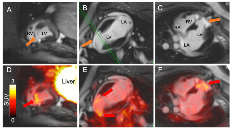
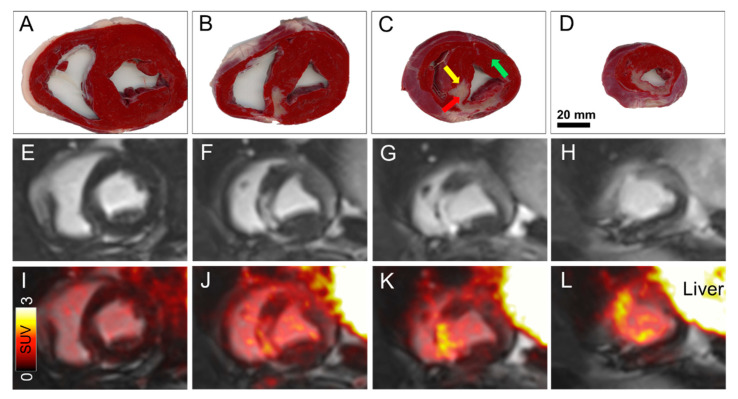
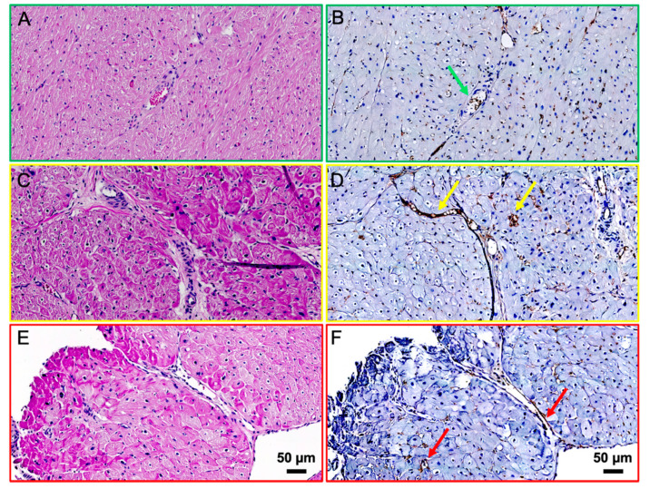
Angiogenesis is crucial in tissue repair and prevents scar tissue formation following an ischemic event such as myocardial infarction. The ischemia induces formation of new capillaries, which have high expression of integrin αvβ3. [68Ga]Ga-NODAGA-E[(cRGDyK)]2 ([68Ga]Ga-RGD) is a promising PET-radiotracer reflecting angiogenesis by binding to integrin αvβ3. A Göttingen mini-pig underwent transient catheter-induced left anterior descending artery (LAD) occlusion for 120 min, and after 8 weeks was imaged on a Siemens mMR 3T PET/MR system. A large antero-septal infarction was evident by late gadolinium enhancement (LGE) on the short axis and 2-4 chamber views. The infarcted area corresponded to the area with high [68Ga]Ga-RGD uptake on the fused PET/MR images, with no uptake in the healthy myocardium. To support the hypothesis that [68Ga]Ga-RGD uptake reflects angiogenesis, biopsies were sampled from the infarct border and healthy myocardium. Expression of αvβ3 was evaluated using immunohistochemistry. The staining showed higher αvβ3 expression in the capillaries of the infarct border compared to those in the healthy myocardium. These initial data confirm in vivo detection of angiogenesis using [68Ga]Ga-RGD PET in a translational model, which overall support the method applicability when evaluating novel cardio-protective therapies.
Keywords: PET/MRI; angiogenesis; cardiac imaging; large animal model; late gadolinium enhancement; magnetic resonance imaging; myocardial infarction; positron emission tomography.
Conflict of interest statement
Andreas Kjaer is listed as the inventor on patents covering the PET tracer used (EP3706809A1 and US16/762,873).
Figures
References
-
- Kiugel M., Dijkgraaf I., Kytö V., Helin S., Liljenbäck H., Saanijoki T., Yim C.-B., Oikonen V., Saukko P., Knuuti G., et al. Dimeric [(68)Ga]DOTA-RGD peptide targeting alphavbeta3 integrin reveals extracellular matrix alterations after myocardial infarction. Mol. Imaging Biol. 2014;16:793–801. doi: 10.1007/s11307-014-0752-1. - DOI - PubMed
-
- Schuleri K.H., Boyle A., Centola M., Amado L.C., Evers R., Zimmet J.M., Evers K.S., Ostbye K.M., Scorpio D.G., Hare J.M., et al. The Adult Gottingen Minipig as a Model for Chronic Heart Failure After Myocardial Infarction: Focus on Cardiovascular Imaging and Regenerative Therapies. Comp. Med. 2008;58:568–579. - PMC - PubMed
-
- Oxboel J., Schjoeth-Eskesen C., El-Ali H.H., Madsen J., Kjaer A. (64)Cu-NODAGA-c(RGDyK) Is a Promising New Angiogenesis PET Tracer: Correlation between Tumor Uptake and Integrin alpha(V)beta(3) Expression in Human Neuroendocrine Tumor Xenografts. Int. J. Mol. Imaging. 2012;2012:379807. doi: 10.1155/2012/379807. - DOI - PMC - PubMed
Grants and funding
LinkOut - more resources
Full Text Sources
