

Keratoconus Diagnostic and Treatment Algorithms Based on Machine-Learning Methods
- PMID: 34679631
- PMCID: PMC8535111
- DOI: 10.3390/diagnostics11101933
Keratoconus Diagnostic and Treatment Algorithms Based on Machine-Learning Methods
Abstract
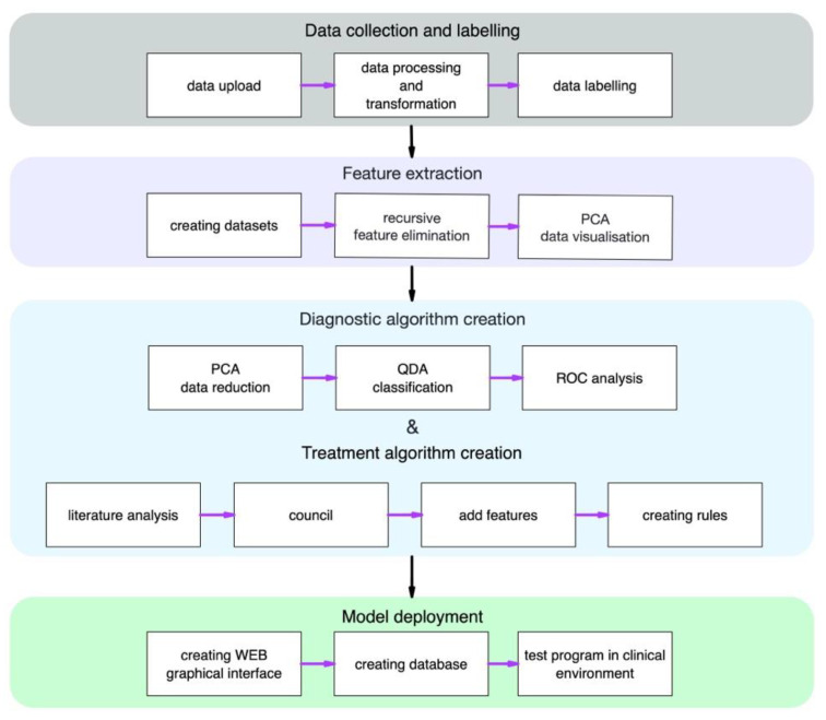
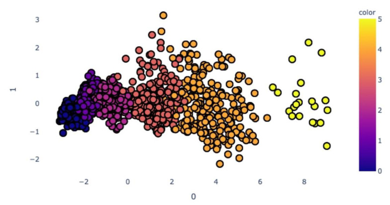
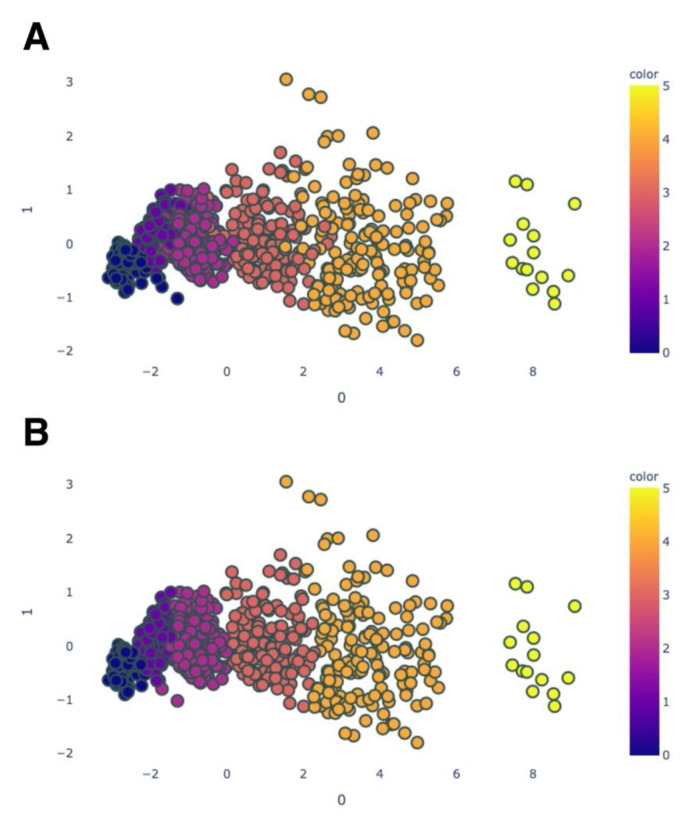
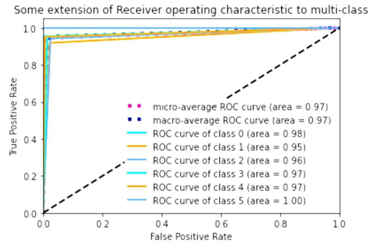
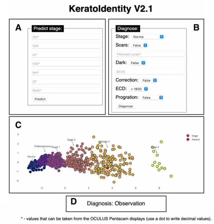
The accurate diagnosis of keratoconus, especially in its early stages of development, allows one to utilise timely and proper treatment strategies for slowing the progression of the disease and provide visual rehabilitation. Various keratometry indices and classifications for quantifying the severity of keratoconus have been developed. Today, many of them involve the use of the latest methods of computer processing and data analysis. The main purpose of this work was to develop a machine-learning-based algorithm to precisely determine the stage of keratoconus, allowing optimal management of patients with this disease. A multicentre retrospective study was carried out to obtain a database of patients with keratoconus and to use machine-learning techniques such as principal component analysis and clustering. The created program allows for us to distinguish between a normal state; preclinical keratoconus; and stages 1, 2, 3 and 4 of the disease, with an accuracy in terms of the AUC of 0.95 to 1.00 based on keratotopographer readings, relative to the adapted Amsler-Krumeich algorithm. The predicted stage and additional diagnostic criteria were then used to create a standardised keratoconus management algorithm. We also developed a web-based interface for the algorithm, providing us the opportunity to use the software in a clinical environment.
Keywords: classification; data visualisation; diagnostics; keratoconus; keratotomography; keratotopography; machine learning; treatment.
Conflict of interest statement
The authors declare no conflict of interest.
Figures
References
-
- George A., Kaufman E.J. StatPearls. StatPearls Publishing; Treasure Island, FL, USA: 2021. [(accessed on 4 August 2021)]. Keratoconus. Available online: http://www.ncbi.nlm.nih.gov/books/NBK470435/
-
- Svetlana I., Komarova O., Semykin A., Zimina M., Konovalova M. Revising the Question of Keratoconus Classification. Int. J. Keratoconus Ectatic Corneal Dis. 2018;7:82–89. doi: 10.5005/jp-journals-10025-1162. - DOI
LinkOut - more resources
Full Text Sources
