. 2021 Oct 6;11(10):1474.
doi: 10.3390/biom11101474.

Activation of Aldehyde Dehydrogenase 2 Ameliorates Glucolipotoxicity of Pancreatic Beta Cells

Affiliations

Activation of Aldehyde Dehydrogenase 2 Ameliorates Glucolipotoxicity of Pancreatic Beta Cells

Shiau-Mei Chen et al. Biomolecules. .

Abstract

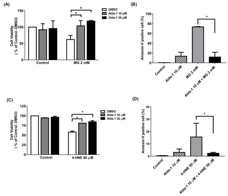
Chronic hyperglycemia and hyperlipidemia hamper beta cell function, leading to glucolipotoxicity. Mitochondrial aldehyde dehydrogenase 2 (ALDH2) detoxifies reactive aldehydes, such as methylglyoxal (MG) and 4-hydroxynonenal (4-HNE), derived from glucose and lipids, respectively. We aimed to investigate whether ALDH2 activators ameliorated beta cell dysfunction and apoptosis induced by glucolipotoxicity, and its potential mechanisms of action. Glucose-stimulated insulin secretion (GSIS) in MIN6 cells and insulin secretion from isolated islets in perifusion experiments were measured. The intracellular ATP concentrations and oxygen consumption rates of MIN6 cells were assessed. Furthermore, the cell viability, apoptosis, and mitochondrial and intracellular reactive oxygen species (ROS) levels were determined. Additionally, the pro-apoptotic, apoptotic, and anti-apoptotic signaling pathways were investigated. We found that Alda-1 enhanced GSIS by improving the mitochondrial function of pancreatic beta cells. Alda-1 rescued MIN6 cells from MG- and 4-HNE-induced beta cell death, apoptosis, mitochondrial dysfunction, and ROS production. However, the above effects of Alda-1 were abolished in Aldh2 knockdown MIN6 cells. In conclusion, we reported that the activator of ALDH2 not only enhanced GSIS, but also ameliorated the glucolipotoxicity of beta cells by reducing both the mitochondrial and intracellular ROS levels, thereby improving mitochondrial function, restoring beta cell function, and protecting beta cells from apoptosis and death.

Keywords: Alda-1; aldehyde dehydrogenase 2 (ALDH2); beta cell function; glucolipotoxicity.


Conflict of interest statement

D.M.-R., C.-H.C., and W.-J.Y. are co-inventors of several issues’ patents on “Modulators of aldehyde dehydrogenase activity and methods of use thereof”, patent Numbers: US 10227304, US 9670162, US 9370506, US 9345693, US 8906942, US 8772295, US 8389522, and US 8354435. W.-J.Y. is an employee and shareholder of Foresee Pharmaceuticals Co. Ltd. W.-J.Y. is a co-inventor of issued patent US 9879036 “Modulators of aldehyde dehydrogenase activity and methods of use thereof”. The other authors declared no competing interests. The funders had no role in the design of the study; in the collection, analyses, or interpretation of data; in the writing of the manuscript, or in the decision to publish the results.

Figures

Figure 1
Figure 1
Alda-1 promotes insulin secretion in MIN6 pancreatic beta cells. (A) Insulin secretory response in MIN6 cells exposed to different doses of Alda-1 under low (3.3 mM) and high glucose (16.7 mM) concentrations (n = 3/group). (B) Glucose-stimulated insulin secretion in ex vivo perifused islets in the absence and presence of 10 μM Alda-1. (C) First and second phase insulin secretions were measured from the islet perifusion study (details in the Method section). (n = 14 in the control group, n = 15 in the Alda-1 10 μM group). Data are presented as mean ± SEM * p < 0.05 versus the control group.
Figure 2
Figure 2
Alda-1 improves mitochondrial function. (A) ATP level in response to 10 μM Alda-1 in MIN6 cells under low (3.3 mM) and high glucose (16.7 mM) concentrations. (B) Effect of Alda-1 on oxygen consumption rate (OCR) in MIN6 beta cells under low (3.3 mM) and high glucose (16.7 mM) concentrations. (C) The area under curves (AUC) of the OCR. Data are presented as mean ± SEM of three independent experiments (n = 3 per group). * p < 0.05 versus the control group with p-for-trend analysis and Student’s t-test, respectively.
Figure 3
Figure 3
Alda-1 rescues the cell death induced by glucotoxicity and lipotoxicity via anti-apoptotic effect. (A) Cell viability in the control and Alda-1-treated MIN6 cells in the absence and presence of 2 mM methylglyoxal (MG). (B) The percentages of Annexin-V-positive and AAD-7-negative MIN6 cells treated with or without Alda-1 in the absence and presence of 2 mM methylglyoxal (MG) for 24 h. (C) Cell viability in control and Alda-1-treated MIN6 cells in the absence and presence of 50 μM 4-Hydroxynonenal (4-HNE). (D) The percentages of the Annexin-V-positive and AAD-7-negative MIN6 cells treated with or without Alda-1 in the absence and presence of 50 μM 4-HNE for 24 h. Data are presented as mean ± SEM of three independent experiments (n = 3 per group). * p < 0.05.
Figure 4
Figure 4
Alda-1 ameliorates mitochondrial dysfunction induced by glucotoxicity and lipotoxicity. The ATP levels in the control and Alda-1-treated MIN6 cells in the absence and presence of 2 mM methylglyoxal (MG) for 24 h (A) or 4-HNE for different time periods (B). Relative mitochondrial (C,E) and cytoplasmic (D,F) ROS levels normalized to MG or 4-HNE in the control and Alda-1-treated MIN6 cells in the absence and presence of 2 mM MG (C,D) or 4-HNE (E,F) for 24 h. (G) The signaling pathway of Alda-1 rescuing 4-HNE-induced cell apoptosis. Treatment of Alda-1 improved the beneficial effects on apoptotic protein levels. Data are presented as mean ± SEM of three independent experiments (n = 3 per group). * p < 0.05.
Figure 5
Figure 5
Aldh2 knockdown abolished the anti-apoptotic effects of ALDH2 activator in MIN6 cells. (A) The knockdown efficiency of shRNA on ALDH2 expression was analyzed by Western blot. Effect of Alda-1 on scramble control and Aldh2 knockdown MIN6 cells on (B) Insulin secretion and (C) ATP levels in low (3.3 mM) and high glucose (16.7 mM) concentrations. Effect of Alda-1 on cell viability in the presence of (D) 2 mM methylglyoxal (MG) or (E) 50 μM 4-HNE. Data are presented as mean ± SEM (n = 3 per group). (F) Incubation in either high glucose or palmitate alone or with both combinations, to evaluate the direct toxic effects of glucolipotoxicity and the protective effect of Alda-1. The data were normalized to 5.5 mM glucose as the control and represented as percentage of viability. (n = 4–6 per group). G: glucose, PA: palmitate. *: p < 0.05. N.S.: not significant.

References

    1. O’Brien P.J., Siraki A.G., Shangari N. Aldehyde sources, metabolism, molecular toxicity mechanisms, and possible effects on human health. Crit. Rev. Toxicol. 2005;35:609–662. doi: 10.1080/10408440591002183. - DOI - PubMed
    1. Robertson R.P., Harmon J., Tran P.O., Poitout V. Beta-cell glucose toxicity, lipotoxicity, and chronic oxidative stress in type 2 diabetes. Diabetes. 2004;53((Suppl. 1)):S119–S124. doi: 10.2337/diabetes.53.2007.S119. - DOI - PubMed
    1. Kohnke R., Mei J., Park M., York D.A., Erlanson-Albertsson C. Fatty acids and glucose in high concentration down-regulates ATP synthase beta-subunit protein expression in INS-1 cells. Nutr. Neurosci. 2007;10:273–278. doi: 10.1080/10284150701745910. - DOI - PubMed
    1. Rhodes C.J. Type 2 diabetes-a matter of beta-cell life and death? Science. 2005;307:380–384. doi: 10.1126/science.1104345. - DOI - PubMed
    1. Thallas-Bonke V., Thorpe S.R., Coughlan M.T., Fukami K., Yap F.Y., Sourris K.C., Penfold S.A., Bach L.A., Cooper M.E., Forbes J.M. Inhibition of NADPH oxidase prevents advanced glycation end product-mediated damage in diabetic nephropathy through a protein kinase C-alpha-dependent pathway. Diabetes. 2008;57:460–469. doi: 10.2337/db07-1119. - DOI - PubMed

Publication types

MeSH terms