. 2021 Oct 18;13(20):5221.
doi: 10.3390/cancers13205221.

Comprehensive Analysis of VEGFR2 Expression in HPV-Positive and -Negative OPSCC Reveals Differing VEGFR2 Expression Patterns

Affiliations

Comprehensive Analysis of VEGFR2 Expression in HPV-Positive and -Negative OPSCC Reveals Differing VEGFR2 Expression Patterns

Senem Uzun et al. Cancers (Basel). .

Abstract

VEGF signaling regulated by the vascular endothelial growth factor receptor 2 (VEGFR2) plays a decisive role in tumor angiogenesis, initiation and progression in several tumors including HNSCC. However, the impact of HPV-status on the expression of VEGFR2 in OPSCC has not yet been investigated, although HPV oncoproteins E6 and E7 induce VEGF-expression. In a series of 56 OPSCC with known HPV-status, VEGFR2 expression patterns were analyzed both in blood vessels from tumor-free and tumor-containing regions and within tumor cells by immunohistochemistry using densitometry. Differences in subcellular colocalization of VEGFR2 with endothelial, tumor and stem cell markers were determined by double-immunofluorescence imaging. Immunohistochemical results were correlated with clinicopathological data. HPV-infection induces significant downregulation of VEGFR2 in cancer cells compared to HPV-negative tumor cells (p = 0.012). However, with respect to blood vessel supply, the intensity of VEGFR2 staining differed only in HPV-positive OPSCC and was upregulated in the blood vessels of tumor-containing regions (p < 0.0001). These results may suggest different routes of VEGFR2 signaling depending on the HPV-status of the OPSCC. While in HPV-positive OPSCC, VEGFR2 might be associated with increased angiogenesis, in HPV-negative tumors, an autocrine loop might regulate tumor cell survival and invasion.

Keywords: cancer stem cell; human papillomavirus; oropharyngeal squamous cell carcinoma; vascular endothelial growth factor receptor 2.


Conflict of interest statement

All other authors declare no conflict of interest.

Figures

Figure 1
Figure 1
Schematic flow diagram of the study design and method. After surgery and tissue processing of OPSCC samples, the HPV-status was determined by GP5+/GP6+ polymerase chain reaction (PCR) and p16INK4A-immunohistochemistry (IHC). To test the specificity of the VEGFR2-antibodies, liver, melanoma, papillary thyroid carcinoma, and cervix squamous cell carcinoma tissues were selected as positive controls. First, hematoxylin and eosin (H&E) staining was performed to determine the regions relevant for further analyses. This was followed by staining of consecutive sections with VEGFR2-AB using the avidin-biotin-peroxidase complex (ABC). Subsequently, densitometric analyses of the VEGFR2-staining intensity of blood vessels in tumor regions and tumor-free regions, as well as tumor cells, were performed. The number of VEGFR2-expressing capillaries in a defined microscopic field was counted and statistically analyzed. Consecutive sections of OPSCC were selected for double immunofluorescence analysis to show colocalization of VEGFR2 and CD31 in endothelial cells (EC), to show the localization of VEGFR2 in tumor cells (TC) by double staining for p16INK4A- (in HPV-positive tumors) or for p53 (in HPV-negative tumors). Confocal double immunofluorescence analysis was performed for VEGFR2 and the stem cell marker ALDH1A1.
Figure 2
Figure 2
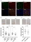
(A) Colocalization analysis of VEGFR2 with CD31 (endothelial cell marker) by immunofluorescence labelling of OPSCC. Antibody stainings were visualized by confocal microscopy. Colocalization of CD31 (a) with VEGFR2 (b) revealed that VEGFR2 is expressed in numerous capillaries of HPV-positive OPSCC (d). Colocalization of CD31 (e) with VEGFR2 (f) revealed that VEGFR2 is only occasionally present in capillaries of HPV-negative tumors (h). Cell nuclei were stained with DRAQ5 (c,g). Scale bar: A-H 20 µm. (B) Representative immunohistochemical staining and corresponding staining intensity analysis of VEGFR2 expression in blood vessels of tumor containing and adjacent tumor-free regions in (left) HPV-positive and (right) HPV-negative OPSCC. HPV-positive tumor regions (M = 0.657 DU; SD = 0.178 DU), adjacent tumor-free regions (M = 0.497; SD = 0.187), (***, p < 0.0001); HPV-negative tumor regions (M = 0.675; SD = 0.251), tumor-free regions (M = 0.616; SD = 0.286), (ns, p = 0.107); HPV-positive and -negative tumor regions (ns, p = 0.740); HPV-positive and-negative tumor-free regions (ns, p = 0.129). (C) Blood vessel density per viewing field of VEGFR2-immunoreactive capillaries in HPV-positive and HPV-negative OPSCC. Vascular density of HPV-positive (M = 45.51; SD = 13.574) and HPV-negative OPSCC (M = 38.85; SD = 14.901), (ns, p = 0.097). (DU = densitometrical units, M = mean, SD = standard deviation, ns = not significant, scale bars: A 50 μm; B 100 μm).
Figure 3
Figure 3
Analysis of VEGFR2 staining intensity in tumor cells of HPV-positive and HPV-negative OPSCC. Representative immunohistochemical VEGFR2 staining images (A = low, B = medium and C = high staining intensity), as well as corresponding H&E stain images of consecutive sections. (A) Staining intensities of low VEGFR2 expressing HPV-positive (M = 0.172 DU; SD = 0.072 DU) and HPV-negative (M = 0.201 DU; SD = 0.067 DU) tumor cells, p = 0.140. (B) Moderate expression levels of VEGFR2-positive tumor cells in HPV-positive (M = 0.478; SD = 0.152) vs. HPV-negative samples (M = 0.600; SD = 0.199), *, p = 0.014. (C) High expression levels of VEGFR2-positive tumor cells in HPV-negative (M = 1.714; SD = 0.260) and HPV-positive specimens (M = 1.382; SD = 0.320), *, p = 0.012. (DU = densitometrical units, M = mean, SD = standard deviation, ns = not significant, scale bar 50 μm).
Figure 4
Figure 4
(A) Colocalization analysis of VEGFR2 with p16INK4A (tumor cell marker of HPV-positive tumors) and VEGFR2 with p53 (tumor cell marker of HPV-negative tumors) by immunofluorescence labeling of OPSCC. Antibody stainings were visualized by confocal microscopy. Colocalization of p16INK4A (a) with VEGFR2 (b) was detected in the cytoplasm and cell nuclei of HPV-positive tumor cells (d). Colocalization of p53 (e) with VEGFR2 (f) was identified in the cytoplasm and cell nuclei of HPV-negative tumor cells. Note, that VEGFR2 was detected in numerous tumor cell nuclei of HPV-positive OPSCC (d) compared to HPV-negative OPSCC (h). Tumor cell nuclei were stained with DRAQ5 (c,g). Scale bar: 20 μm. (B) Colocalization analysis of VEGFR2 with ALDH1A1 (CSC marker) by immunofluorescence labeling of OPSCC. Immunohistochemical staining against VEGFR2 of a representative consecutive HPV-positive (af) and HPV-negative tumor section (gl). (a,g) Overview and (b,h) details. A subpopulation of tumor cells with strong VEGFR2 immunoreactivity can be observed. Colocalization of VEGFR2 (d,j) with ALDH1A1 (f,l) was detected only in a subpopulation of tumor cells and at the subcellular level mainly in the cytoplasm (asterisks). The cells were distributed around the blood vessels (f,l). In HPV-positive OPSCC, some migrating cells were detected at the blood vessel wall and one cell is visible intravasally, while others were recognized in the fibrous tissue (f). Cell nuclei were stained with DRAQ5 (e,k). Scale bars: (a,g) 200 μm; (b,h) 50 μm; (cf), (il) 20 μm.
Figure 5
Figure 5
Schematic presentation of the two suggested HPV-status-dependent VEGFR2 signal pathways in OPSCC. Depending on HPV and possibly triggered by hypoxia and/or oxidative stress, VEGFR2 expression is upregulated in tumor blood vessels of HPV-positive OPSCC, which may be associated with increased angiogenesis. This is not observed in HPV-negative OPSCC; instead, VEGFR2 is significantly stronger expressed in the tumor cells themselves, which may lead to increased activation of tumor cell proliferation, migration, invasion and reduced apoptosis. The increased tumor cell activity may also be correlated to the tendency of lower blood vessel count in HPV-negative OPSCC due to hypoxia. Figure modified from [10].

References

    1. Chaturvedi A.K., Engels E.A., Pfeiffer R.M., Hernandez B.Y., Xiao W., Kim E., Jiang B., Goodman M.T., Sibug-Saber M., Cozen W., et al. Human papillomavirus and rising oropharyngeal cancer incidence in the United States. J. Clin. Oncol. 2011;29:4294–4301. doi: 10.1200/JCO.2011.36.4596. - DOI - PMC - PubMed
    1. CDC Cancers Associated with Human Papillomavirus, United States—2012–2016. USCS Data Brief, no 10. [(accessed on 8 February 2020)]; Available online: https://www.cdc.gov/cancer/uscs/about/data-briefs/no10-hpv-assoc-cancers....
    1. Leemans C.R., Snijders P.J.F., Brakenhoff R.H. The molecular landscape of head and neck cancer. Nat. Rev. Cancer. 2018;18:269–282. doi: 10.1038/nrc.2018.11. - DOI - PubMed
    1. Hoffmann M., Tribius S. HPV and oropharyngeal squamous cell cancer in the 8th edition of the TNM classification. Laryngorhinootologie. 2018;97:641–649. doi: 10.1055/s-0043-123028. - DOI - PubMed
    1. Simons M., Gordon E., Claesson-Welsh L. Mechanisms and regulation of endothelial VEGF receptor signalling. Nat. Rev. Molec. Cell Biol. 2016;17:611–625. doi: 10.1038/nrm.2016.87. - DOI - PubMed

LinkOut - more resources