. 2021 Oct 18;13(20):5228.
doi: 10.3390/cancers13205228.

Combining Hypermethylated RASSF1A Detection Using ddPCR with miR-371a-3p Testing: An Improved Panel of Liquid Biopsy Biomarkers for Testicular Germ Cell Tumor Patients

Affiliations

Combining Hypermethylated RASSF1A Detection Using ddPCR with miR-371a-3p Testing: An Improved Panel of Liquid Biopsy Biomarkers for Testicular Germ Cell Tumor Patients

João Lobo et al. Cancers (Basel). .

Abstract

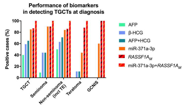
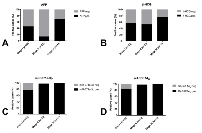
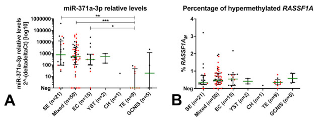
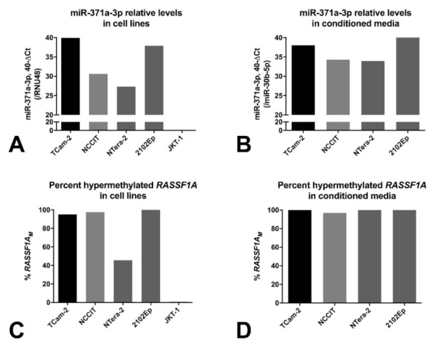
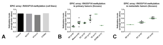
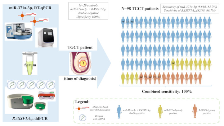
The classical serum tumor markers used routinely in the management of testicular germ cell tumor (TGCT) patients-alpha fetoprotein (AFP) and human chorionic gonadotropin (HCG)-show important limitations. miR-371a-3p is the most recent promising biomarker for TGCTs, but it is not sufficiently informative for detection of teratoma, which is therapeutically relevant. We aimed to test the feasibility of hypermethylated RASSF1A (RASSF1AM) detected in circulating cell-free DNA as a non-invasive diagnostic marker of testicular germ cell tumors, combined with miR-371a-3p. A total of 109 serum samples of patients and 29 sera of healthy young adult males were included, along with representative cell lines and tumor tissue samples. We describe a novel droplet digital polymerase chain reaction (ddPCR) method for quantitatively assessing RASSF1AM in liquid biopsies. Both miR-371a-3p (sensitivity = 85.7%) and RASSF1AM (sensitivity = 86.7%) outperformed the combination of AFP and HCG (sensitivity = 65.5%) for TGCT diagnosis. RASSF1AM detected 88% of teratomas. In this representative cohort, 14 cases were negative for miR-371a-3p, all of which were detected by RASSF1AM, resulting in a combined sensitivity of 100%. We have described a highly sensitive and specific panel of biomarkers for TGCT patients, to be validated in the context of patient follow-up and detection of minimal residual disease.

Keywords: RASSF1A; biomarkers; droplet digital PCR; germ cell tumors; methylation; miR-371a-3p; microRNAs.


Conflict of interest statement

The authors declare no conflict of interest.

Figures

Figure 1
Figure 1
Performance of AFP, β-HCG, miR-371a-3p and RASSF1AM in detecting testicular germ cell tumor patients at time of diagnosis. Results are presented color-coded, referring to all testicular germ cell tumor patients (n = 98); all seminomas (n = 21); all non-seminomas (including teratomas, n = 77); all teratomas (n = 9); and all GCNIS-only patients (n = 5). Notice that both miR-371a-3p and RASSF1AM show better detection performance than the combination of both AFP and β-HCG, and that combination of the former two biomarkers results in 100% sensitivity for detecting TGCT patients, including those with teratoma and those with GCNIS-only. Abbreviations: AFP—alpha fetoprotein; β-HCG—human chorionic gonadotropin subunit β; GCNIS—germ cell neoplasia in situ; RASSF1AM—hypermethylated RASSF1A; TE—teratoma.
Figure 2
Figure 2
Proportion of positive cases for AFP (A), β-HCG (B), miR-371a-3p (C) and RASSF1AM (D) according to stage of disease. Abbreviations: AFP—alpha fetoprotein; β-HCG—human chorionic gonadotropin subunit β; RASSF1AM—hypermethylated RASSF1A.
Figure 3
Figure 3
Relative levels of miR-371a-3p (A) and percentage of RASSF1AM (B) across histological subtypes. Negative samples (as by the defined cutoffs) are plotted at the xx-axis (Neg). Stage I patients are highlighted in red. miR-371a-3p relative levels are normalized to miR-30b-5p and plotted in log10 format for readability. Bars represent median and interquartile range. * p < 0.05; ** p < 0.01; *** p < 0.001 (adjusted for multiple comparisons). Abbreviations: CH—choriocarcinoma; EC—embryonal carcinoma; GCNIS—germ cell neoplasia in situ; SE—seminoma; RASSF1AM—hypermethylated RASSF1A; TE—teratoma; YST—yolk sac tumor.
Figure 4
Figure 4
Relative levels of miR-371a-3p in cell lines (A) and in conditioned media (B), and percentage of RASSF1AM in cell lines (C) and conditioned media (D). miR-371a-3p relative levels are plotted in 40-ΔCt format for readability (normalized to RNU48 or miR-30b-5p in cell lines and conditioned media, respectively). Abbreviations: RASSF1AM—hypermethylated RASSF1A.
Figure 5
Figure 5
Estimated methylation levels for RASSF1A promoter derived from EPIC array analyses on cell lines (A), primary tumor tissues (B) and metastatic tumor tissues (C). Average beta values are plotted. Notice that the single teratoma sample has the highest methylation levels. Abbreviations: EC—embryonal carcinoma; GCNIS—germ cell neoplasia in situ; SE—seminoma; TE—teratoma; YST—yolk sac tumor.
Figure 6
Figure 6
Illustrative overview of the combined pipeline. Serum samples of 98 TGCT patients and 29 healthy male blood donors were submitted to miR-371a-3p quantification by real-time quantitative PCR and to RASSF1AM quantification via ddPCR. Each male patient figure represents one of the TGCT patients in the study. The color code identifies if the patient was diagnosed because of positivity for both markers (blue), positivity for miR-371a-3p only (yellow) or positivity for RASSF1A only (orange). The patients with pure teratoma are highlighted by “TE”. The combined pipeline detected all TGCT patients (including those with pure teratoma) while all tested controls were negative, resulting in a combined sensitivity and specificity of 100%. Abbreviations: ddPCR—droplet digital PCR; cfDNA—cell-free DNA; RASSF1AM—hypermethylated RASSF1A; RT-qPCR—real-time quantitative PCR; TE—teratoma; TGCT—testicular germ cell tumor.

References

    1. Wan J.C.M., Massie C., Garcia-Corbacho J., Mouliere F., Brenton J.D., Caldas C., Pacey S., Baird R., Rosenfeld N. Liquid biopsies come of age: Towards implementation of circulating tumour DNA. Nat. Rev. Cancer. 2017;17:223–238. doi: 10.1038/nrc.2017.7. - DOI - PubMed
    1. Siravegna G., Mussolin B., Venesio T., Marsoni S., Seoane J., Dive C., Papadopoulos N., Kopetz S., Corcoran R.B., Siu L.L., et al. How liquid biopsies can change clinical practice in oncology. Ann. Oncol. 2019;30:1580–1590. doi: 10.1093/annonc/mdz227. - DOI - PubMed
    1. Rossi G., Ignatiadis M. Promises and Pitfalls of Using Liquid Biopsy for Precision Medicine. Cancer Res. 2019;79:2798–2804. doi: 10.1158/0008-5472.CAN-18-3402. - DOI - PubMed
    1. Gai W., Sun K. Epigenetic Biomarkers in Cell-Free DNA and Applications in Liquid Biopsy. Genes. 2019;10:32. doi: 10.3390/genes10010032. - DOI - PMC - PubMed
    1. Locke W.J., Guanzon D., Ma C., Liew Y.J., Duesing K.R., Fung K.Y.C., Ross J.P. DNA Methylation Cancer Biomarkers: Translation to the Clinic. Front. Genet. 2019;10:1150. doi: 10.3389/fgene.2019.01150. - DOI - PMC - PubMed

LinkOut - more resources