Review
. 2021 Oct 1;9(10):1366.
doi: 10.3390/biomedicines9101366.

Inflammation in Duchenne Muscular Dystrophy-Exploring the Role of Neutrophils in Muscle Damage and Regeneration

Affiliations
Review

Inflammation in Duchenne Muscular Dystrophy-Exploring the Role of Neutrophils in Muscle Damage and Regeneration

Ankita Tulangekar et al. Biomedicines. .

Abstract

Duchenne muscular dystrophy (DMD) is a severe and progressive, X-linked, neuromuscular disorder caused by mutations in the dystrophin gene. In DMD, the lack of functional dystrophin protein makes the muscle membrane fragile, leaving the muscle fibers prone to damage during contraction. Muscle degeneration in DMD patients is closely associated with a prolonged inflammatory response, and while this is important to stimulate regeneration, inflammation is also thought to exacerbate muscle damage. Neutrophils are one of the first immune cells to be recruited to the damaged muscle and are the first line of defense during tissue injury or infection. Neutrophils can promote inflammation by releasing pro-inflammatory cytokines and compounds, including myeloperoxidase (MPO) and neutrophil elastase (NE), that lead to oxidative stress and are thought to have a role in prolonging inflammation in DMD. In this review, we provide an overview of the roles of the innate immune response, with particular focus on mechanisms used by neutrophils to exacerbate muscle damage and impair regeneration in DMD.

Keywords: DMD; Duchenne muscular dystrophy; inflammation; myeloperoxidase; neutrophil elastase; neutrophils.


Conflict of interest statement

The authors declare no conflict of interest.

Figures

Figure 1
Figure 1
Schematic of the immunological events following muscle damage in Duchenne muscular dystrophy (DMD). An inflammatory response is activated in dystrophic muscle cells, and immune cells, including neutrophils and macrophages, are recruited to the sites of damage. The expression of inducible nitric oxide synthase (iNOS), myeloperoxidase (MPO), hypochlorous acid (HOCl), and pro-inflammatory cytokines, including interleukin (IL) 6 (IL-6), tumor necrosis factor alpha (TNF-α) and IL-1β, followed by anti-inflammatory cytokines, including IL-10, IL-4 and transforming growth factor beta (TGF-β), combined with the release of DAMPs including single stranded RNA (ssRNA) and high mobility group box protein 1 (HMGB1), initially results in regeneration of the muscle. However, continuous release of cytokines and DAMPs results in prolonged inflammation. This chronic inflammatory condition leads to impaired muscle repair followed by necrosis of muscle cells and accumulation of excessive fatty connective tissue leading to fibrosis.
Figure 2
Figure 2
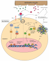
Mechanisms used by neutrophils to promote muscle damage in Duchenne muscular dystrophy (DMD). Following muscle damage, damage associated molecular patterns (DAMPS) are released from the dystrophic muscle and activate neutrophils via recognition by toll-like receptors (TLRs) and macrophage-1 antigen (Mac-1) on the cell surface. This interaction activates the myeloid differentiation primary response 88 (MyD88) pathway which further activates the IκB kinases (IKKs) and mitogen-activated kinases (MAPKs). This induces the expression of nuclear factor kappa B (NF-κB) and activator protein 1 (AP-1) transcription factors which promote the transcription of pro-inflammatory cytokines. DAMP-TLR interactions also cause the release of neutrophil elastase (NE) and myeloperoxidase (MPO) from the azurophilic granules within the neutrophil into the cytoplasm. MPO catalyzes the production of reactive oxygen species (ROS) including hypochlorous acid (HOCl), which elevates oxidative stress and promotes muscle cell lysis. NE induces chromatin decondensation and, together with MPO, lead to neutrophil extracellular trap (NET) formation. It is thought that NETs are released outside the cell by cell-lysis and further promote inflammation.

References

    1. Blake D.J., Weir A., Newey S.E., Davies K.E. Function and genetics of dystrophin and dystrophin-related proteins in muscle. Physiol. Rev. 2002;82:291–329. doi: 10.1152/physrev.00028.2001. - DOI - PubMed
    1. Omairi S., Hau K.-L., Collins-Hooper H., Scott C., Vaiyapuri S., Torelli S., Montanaro F., Matsakas A., Patel K. Regulation of the dystrophin-associated glycoprotein complex composition by the metabolic properties of muscle fibres. Sci. Rep. 2019;9:2770. doi: 10.1038/s41598-019-39532-4. - DOI - PMC - PubMed
    1. Tidball J.G., Welc S.S., Wehling-Henricks M. Immunobiology of inherited muscular dystrophies. Compr. Physiol. 2018;8:1313–1356. doi: 10.1002/cphy.c170052. - DOI - PMC - PubMed
    1. Nowak K.J., Davies K.E. Duchenne muscular dystrophy and dystrophin: Pathogenesis and opportunities for treatment. EMBO Rep. 2004;5:872–876. doi: 10.1038/sj.embor.7400221. - DOI - PMC - PubMed
    1. Grounds M.D., Terrill J.R., Al-Mshhdani B.A., Duong M.N., Radley-Crabb H.G., Arthur P.G. Biomarkers for Duchenne muscular dystrophy: Myonecrosis, inflammation and oxidative stress. Dis. Model. Mech. 2020;13:dmm043638. doi: 10.1242/dmm.043638. - DOI - PMC - PubMed