. 2021 Oct 5;9(10):1395.
doi: 10.3390/biomedicines9101395.

Protein Unfolding: Denaturant vs. Force

Affiliations

Protein Unfolding: Denaturant vs. Force

Colleen Kelly et al. Biomedicines. .

Abstract

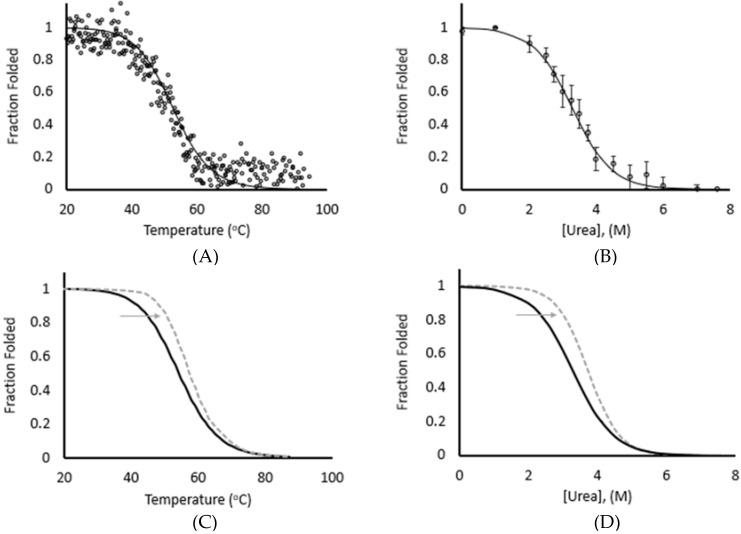
While protein refolding has been studied for over 50 years since the pioneering work of Christian Anfinsen, there have been a limited number of studies correlating results between chemical, thermal, and mechanical unfolding. The limited knowledge of the relationship between these processes makes it challenging to compare results between studies if different refolding methods were applied. Our current work compares the energetic barriers and folding rates derived from chemical, thermal, and mechanical experiments using an immunoglobulin-like domain from the muscle protein titin as a model system. This domain, I83, has high solubility and low stability relative to other Ig domains in titin, though its stability can be modulated by calcium. Our experiments demonstrated that the free energy of refolding was equivalent with all three techniques, but the refolding rates exhibited differences, with mechanical refolding having slightly faster rates. This suggests that results from equilibrium-based measurements can be compared directly but care should be given comparing refolding kinetics derived from refolding experiments that used different unfolding methods.

Keywords: chemical denaturation; immunoglobulin domain; magnetic tweezers; protein refolding; thermal denaturation; titin.


Conflict of interest statement

The authors declare no conflict of interest.

Figures

Figure 1
Figure 1
Thermal and chemical unfolding of I83 yield similar free energies and calcium stabilization. (A) CD data obtained by thermal melt for I83 (○) is fit by a two-state unfolding model (solid line) with a thermal midpoint (Tm) of 53.2 °C. (B) Center of mass calculated from tryptophan fluorescence spectra measured during chemical unfolding (○) is fit by a two-state unfolding model, with a midpoint ([Urea]50%) of 3.2 M. Error bars show ± one standard deviation. (C) Thermal unfolding fit of I83 in the absence of calcium (black) is overlayed with the unfolding fit at pCa 4.3 (dashed gray line). (D) Chemical unfolding fit of I83 in the absence of calcium (black) is overlayed with the unfolding fit at pCa 4.3 (dashed gray line). Reprinted from Reference [4].
Figure 2
Figure 2
Step-fitting of 35-pN unfolding of (I83)6 polymer. Unfolding data demonstrated system noise of approximately ± 20 nm (light gray). A moving average (dark gray) and Kerssemaker fit (black line) more clearly illustrate the extension, unfolding, and refolding of the hexamer. The applied force over time, shown below the data trace, started at 4 pN, was stepped to 35 pN, and then returned to 4 pN after ~45 s. The unfolding steps of the individual Ig domains are marked by triangles and follow the initial elastic extension of the polymer.
Figure 3
Figure 3
Sample unfolding plots from magnetic tweezers exhibit slower unfolding rates in presence of calcium. (A) Unfolding steps at 40 pN occurred more rapidly in the absence of calcium (black) than in the presence of calcium (dashed gray). (B) Unfolding steps at 50 pN occurred slightly more rapidly in the absence of calcium (black) than in the presence of calcium (dashed gray). The first unfolding step is not visible when unfolding at 50 pN because it occurs during the elastic extension step.
Figure 4
Figure 4
Sample refolding traces of (I83)6 polymer in calcium shifts equilibrium to folded state. The same molecule was monitored in absence (black) and presence (gray) of calcium. A histogram is used to show the distribution of the number of folded domains as measured by extension values over 60 s. A shift toward the folded state is observed in the presence of calcium at (A) 4 pN, (B) 6 pN, (C) 8 pN, and (D) 10 pN.
Figure 5
Figure 5
Refolding Traces of (I83)6 polymer at 4 pN and 10 pN. Sample unfolding traces at (A) 4 pN and (B) 10 pN of force illustrate faster and more dynamic refolding in presence of calcium (dashed gray) vs. the absence of calcium (black) as demonstrated by a greater decrease in extension and more frequent folding transitions over time.
Figure 6
Figure 6
Chevron plots for I83 yield similar, but different rate constants at zero denaturant and zero force. (A) The natural log of unfolding rate constants for urea denaturation yielded slower unfolding at pCa 4.3. Average values (N = 4) in the absence of calcium (black) and in the presence of calcium (gray) are shown ± one standard deviation. (B) The natural log of force-induced unfolding and refolding rates illustrate slower unfolding between 30 and 40 pN and fast refolding at force ≤10 pN. Average values (N = 5) in the absence of calcium (black) and in the presence of calcium (gray) are shown ± one standard deviation.

References

    1. Anfinsen C.B. Principles that govern the folding of protein chains. Science. 1973;181:223–230. doi: 10.1126/science.181.4096.223. - DOI - PubMed
    1. Levinthal C. Are there pathways for protein folding? J. Chim. Phys. 1968;65:44–45. doi: 10.1051/jcp/1968650044. - DOI
    1. Eftink M.R. The use of fluorescence methods to monitor unfolding transitions in proteins. Biophys. J. 1994;66:482–501. doi: 10.1016/S0006-3495(94)80799-4. - DOI - PMC - PubMed
    1. Kelly C., Pace N., Gage M., Pfuhl M. Solution NMR Structure of Titin N2A Region Ig Domain I83 and Its Interaction with Metal Ions. J. Mol. Biol. 2021;433:166977. doi: 10.1016/j.jmb.2021.166977. - DOI - PMC - PubMed
    1. Royer C.A. Probing Protein Folding and Conformational Transitions with Fluorescence. Chem. Rev. 2006;106:1769–1784. doi: 10.1021/cr0404390. - DOI - PubMed

LinkOut - more resources