. 2021 Oct 9;9(10):1419.
doi: 10.3390/biomedicines9101419.

Initial Biological Assessment of Upconversion Nanohybrids

Affiliations

Initial Biological Assessment of Upconversion Nanohybrids

Juan Ferrera-González et al. Biomedicines. .

Abstract

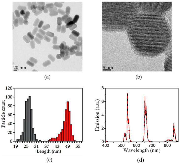
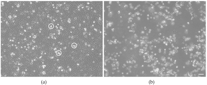
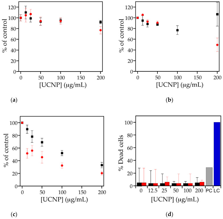
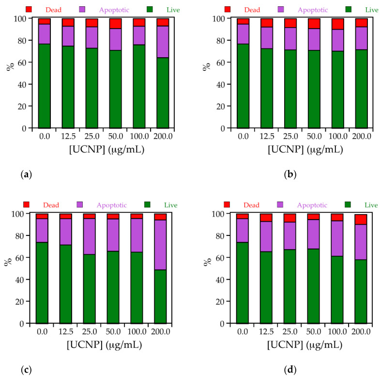
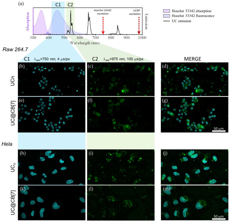
Nanoparticles for medical use should be non-cytotoxic and free of bacterial contamination. Upconversion nanoparticles (UCNPs) coated with cucurbit[7]uril (CB[7]) made by combining UCNPs free of oleic acid, here termed bare UCNPs (UCn), and CB[7], i.e., UC@CB[7] nanohybrids, could be used as photoactive inorganic-organic hybrid scaffolds for biological applications. UCNPs, in general, are not considered to be highly toxic materials, but the release of fluorides and lanthanides upon their dissolution may cause cytotoxicity. To identify potential adverse effects of the nanoparticles, dehydrogenase activity of endothelial cells, exposed to various concentrations of the UCNPs, was determined. Data were verified by measuring lactate dehydrogenase release as the indicator of loss of plasma membrane integrity, which indicates necrotic cell death. This assay, in combination with calcein AM/Ethidium homodimer-1 staining, identified induction of apoptosis as main mode of cell death for both particles. The data showed that the UCNPs are not cytotoxic to endothelial cells, and the samples did not contain endotoxin contamination. Higher cytotoxicity, however, was seen in HeLa and RAW 264.7 cells. This may be explained by differences in lysosome content and particle uptake rate. Internalization of UCn and UC@CB[7] nanohybrids by cells was demonstrated by NIR laser scanning microscopy.

Keywords: cucurbituril; cytotoxicity; upconversion nanoparticles.


Conflict of interest statement

The authors declare no conflict of interest.

Figures

Figure 1
Figure 1
Representative HRTEM images of (a) UCn and, (b) UC@CB[7] nanoparticles. (c) Histogram showing the size distribution of the NPs for the width (grey bars) and length (red bars). (d) Corrected emission spectra (λex = 975 nm) of 10 mg/mL DMSO dispersions of ligand-free UCNPs (black line) and UC@CB[7] (red dotted line).
Figure 2
Figure 2
Morphology of untreated EAhy 926 endothelial cells (a) and cells exposed to the positive control 20 nm plain polystyrene particles (b) after 24 h of culture. Cultures of the untreated EAhy 926 endothelial cells have formed a confluent monolayer with occasional dividing cells (indicated by circles). Cells exposed to the positive control do not form a monolayer but have rounded up, detached from the growth substrate, and float in the medium. Scale bar: 50 µm.
Figure 3
Figure 3
Cell viability (%) of (a) EAhy926 endothelial cells, (b) HeLa, and (c) RAW 264.7 cells according to dehydrogenase activity after incubation with different concentrations of UC@CB[7] (black squares) and UCn (red circles). Cells without exposure to nanoparticles represent 100%. (d) LDH release after exposure of EAhy926 cells to different concentrations of UCNPs for 24h. UC@CB[7] (black bar), UCn (red bar), positive control (PC, grey bar), and lysis control (LC, blue bar). Values are shown as mean ± standard deviation.
Figure 4
Figure 4
Stack column graph representing the percentages of live (green), dead (red), and apoptotic (purple) cells for treated (top) HeLa and (bottom) RAW 264.7 cells in the presence of 0, 12.5, 25.0, 50.0, 100.0, and 200.0 μg/mL of (a and c, respectively) UCn or (b and d, respectively) UC@CB[7].
Figure 5
Figure 5
Morphology of (left) HeLa and (right) RAW 264.7 cells exposed to 50 µg/mL after 24 h of culture with untreated, UCn and UC@CB[7] nanoparticles (a,c,e and b,d,f, respectively).
Figure 6
Figure 6
(a) Absorption (pink area) and fluorescence (purple area) of Hoechst 33342, emission of UCn dispersion in DMSO (black line) together with the wavelength used to excite the dye/UCNP (arrows) and the microscope detection channels (coloured rectangles). (bg) microscope images of RAW 264.7 cells exposed to UCn and UC@CB[7]. (hm) microscope images of HeLa cells exposed to UCn and UC@CB[7]. Left column shows the fluorescence signal of Hoechst 33342 (λex = 750 nm, dwell time: 4 μs/pixel, detection channel: C1) central column shows the 520/540 nm Er3+ upconversion emission (λex = 975 nm, dwell time: 100 μs/pixel, detection channel: C2) and right column is a composite of the in line previous images. 50 μm scale bar applies for all images.

References

    1. Francés-Soriano L., González-Béjar M., Pérez-Prieto J. Synergistic Effects in organic-coated upconversion nanoparticles. In: Altavilla C., editor. Upconverting Nanomaterials: Perspectives, Synthesis, and Applications. CRC Press; Boca Raton, FL, USA: 2016. pp. 101–138.
    1. Li Z., Li X., Yang Y.-W. Photoactive nanoparticles capped with macrocycles as platforms and hosts. In: Pérez-Prieto J., González-Béjar M., editors. Micro and Nano Technologies. Elsevier; Amsterdam, The Netherlands: 2019. pp. 139–167.
    1. Ramasamy T., Ruttala H.B., Gupta B., Poudel B.K., Choi H.-G., Yong C.S., Kim J.O. Smart chemistry-based nanosized drug delivery systems for systemic applications: A comprehensive review. J. Control. Release. 2017;258:226–253. doi: 10.1016/j.jconrel.2017.04.043. - DOI - PubMed
    1. Ramasamy T., Munusamy S., Ruttala H.B., Kim J.O. Smart Nanocarriers for the delivery of nucleic acid-based therapeutics: A comprehensive review. Biotechnol. J. 2021;16:1900408. doi: 10.1002/biot.201900408. - DOI - PubMed
    1. González-Béjar M., Francés-Soriano L., Pérez-Prieto J. Upconversion nanoparticles for bioimaging and regenerative medicine. Front. Bioeng. Biotechnol. 2016;4:47. doi: 10.3389/fbioe.2016.00047. - DOI - PMC - PubMed

LinkOut - more resources